. 2002 Sep 1;22(17):7737-45.
doi: 10.1523/JNEUROSCI.22-17-07737.2002.

Phosphorylation of extracellular signal-regulated kinase in primary afferent neurons by noxious stimuli and its involvement in peripheral sensitization

Affiliations

Phosphorylation of extracellular signal-regulated kinase in primary afferent neurons by noxious stimuli and its involvement in peripheral sensitization

Yi Dai et al. J Neurosci. .

Abstract

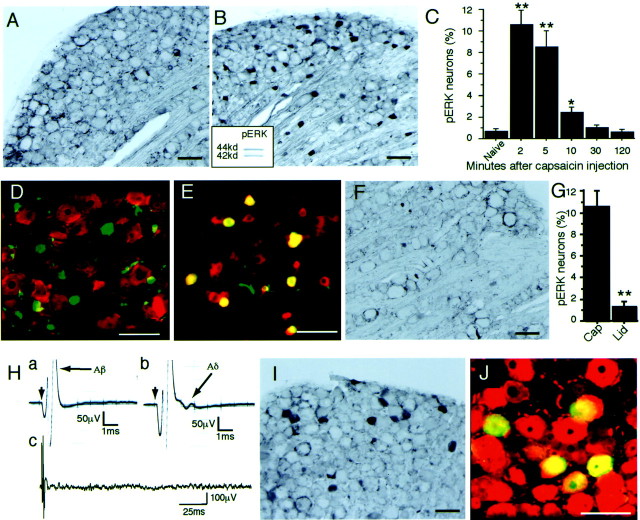
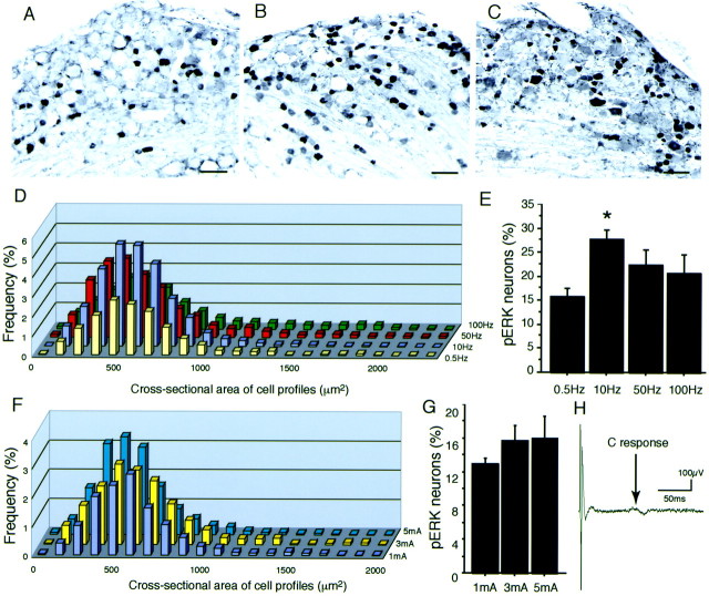
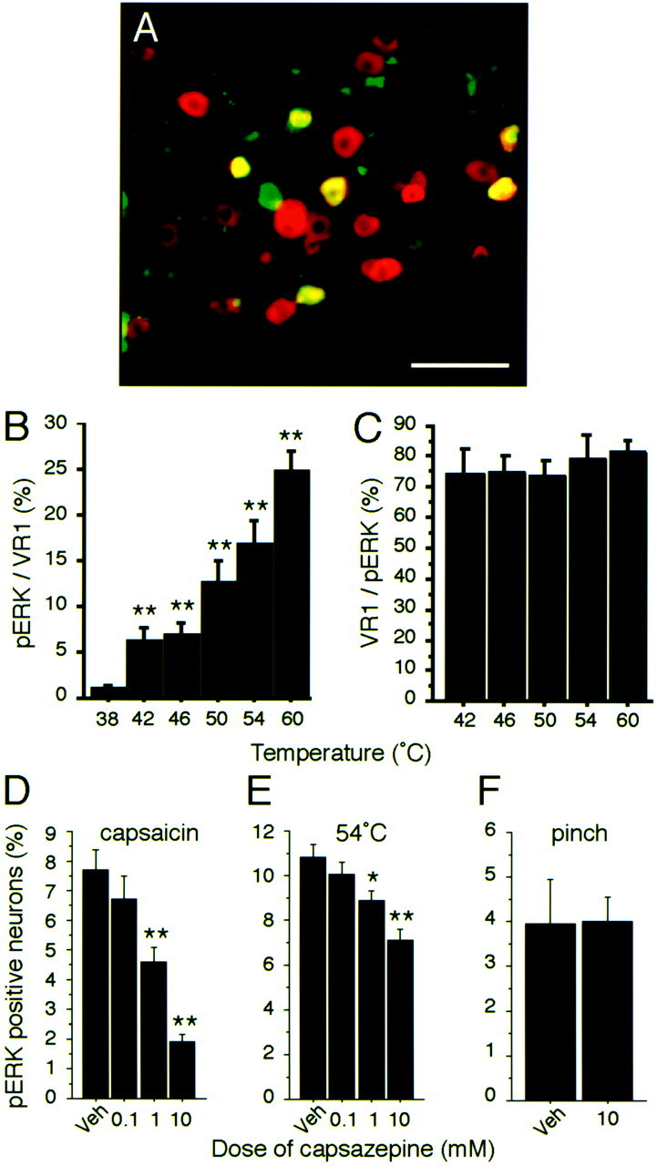
Alteration in the intracellular signal transduction pathway in primary afferent neurons may contribute to pain hypersensitivity. We demonstrated that very rapid phosphorylation of extracellular signal-regulated protein kinases (pERK) occurred in DRG neurons that were taking part in the transmission of various noxious signals. The electrical stimulation of Adelta fibers induced pERK primarily in neurons with myelinated fibers. c-Fiber activation by capsaicin injection induced pERK in small neurons with unmyelinated fibers containing vanilloid receptor-1 (VR-1), suggesting that pERK labeling in DRG neurons is modality specific. Electrical stimulation at the c-fiber level with different intensities and frequencies revealed that phosphorylation of ERK is dependent on the frequency. We examined the pERK in the DRG after application of natural noxious stimuli and found a stimulus intensity-dependent increase in labeled cell size and in the number of activated neurons in the c- and Adelta-fiber population. Immunohistochemical double labeling with phosphorylated ERK/VR-1 and pharmacological study demonstrated that noxious heat stimulation induced pERK in primary afferents in a VR-1-dependent manner. Capsaicin injection into the skin also increased pERK labeling significantly in peripheral fibers and terminals in the skin, which was prevented by a mitogen-activated protein kinase/ERK kinase inhibitor, 1,4-diamino-2,3-dicyano-1,4-bis(2-aminopheylthio)butadiene (U0126). Behavioral experiments showed that U0126 dose-dependently attenuated thermal hyperalgesia after capsaicin injection and suggested that the activation of ERK pathways in primary afferent neurons is involved in the sensitization of primary afferent neurons. Thus, pERK in primary afferents by noxious stimulation in vivo showed distinct characteristics of expression and may be correlated with the functional activity of primary afferent neurons.


Figures

Fig. 1.
Fig. 1.
Stimulus-evoked ERK phosphorylation in rat DRG neurons. A, L4 DRG section (30 μm) immunostained for pERK in the naive rat. B, pERK labeling in many small L4 DRG neurons 2 min after intraplantar injection of capsaicin (10 mm, 200 μl). The inset shows a Western blot of capsaicin-stimulated DRG tissue with pERK antibody.C, Time course of capsaicin-evoked pERK expression in L4/5 DRG neurons (n = 5 at each time point; *p < 0.01; **p < 0.001 compared with naive). D, L4 DRG section double-immunostained for NF200 (red) and pERK (green) after intraplantar capsaicin injection.E, Colocalization of pERK (green) and VR-1 (red) in L4 DRG neurons after capsaicin injection. Double staining appears yellow. F, G, Capsaicin-induced pERK was inhibited by the lidocaine block to the sciatic nerve. Cap, Capsaicin group;Lid, capsaicin plus lidocaine group (**p < 0.001 compared with capsaicin group).H, Example of 0.1 mA (a) and 0.3 mA (b, c) evoked action potentials at the sciatic nerve.a, Aβ response; b, c, Aβ and Aδ responses with no c response (c; 100 sweeps were averaged). I, L4 DRG section immunostained for pERK 2 min after 0.3 mA of stimulation to the sciatic nerve. J, L4 DRG section immunostained for NF200 (a marker of myelinated neurons,red) and pERK (green) after a 0.3 mA electrical stimulation. Double staining appearsyellow. Scale bars, 100 μm.
Fig. 2.
Fig. 2.
Electrical stimulation-induced pERK expression.A–C, Photomicrographs of pERK-labeled neurons in L4 DRG 2 min after 60 pulses of 3 mA/0.5 Hz (A), 3 mA/10 Hz (B), and 3 mA/100 Hz (C) electrical stimulation to the sciatic nerve. D, Size distribution of pERK-labeled neurons in the L4/5 DRGs in the rats that received 60 pulses of 3 mA/0.5 Hz, 3 mA/10 Hz, 3 mA/50 Hz, and 3 mA/100 Hz (n = 3 for each frequency) electrical stimulation to the sciatic nerve. E, The percentage of pERK-labeled neurons in L4/5 DRG neurons (n = 3 for each frequency; ANOVA test shows ap < 0.05 significant change among the groups; *p < 0.01 compared with the 0.5 Hz group by Fisher's PLSD test). F, Size distribution of pERK-labeled neurons in the L4/5 DRGs in the rats that received 60 pulses of 1 mA/0.5 Hz, 3 mA/0.5 Hz, and 5 mA/0.5 Hz (n = 3 for each intensity) electrical stimulation to the sciatic nerve. G, The percentage of pERK-labeled neurons in L4/5 DRG neurons (n = 3 for each intensity; no significant change by ANOVA). H, Electrical stimulation (3 mA) to the sciatic nerve showed both A- and C-fiber responses (100 sweeps were averaged). Scale bars, 100 μm.
Fig. 3.
Fig. 3.
Thermal stimulus intensity-dependent pERK expression. A, pERK labeling in L4 DRG neurons 2 min after thermal stimulation at 42°C. B, Colocalization of pERK (green) and PGP 9.5 (red) in L4 DRG neurons after the 42°C stimulus. Double staining appearsyellow. C, pERK labeling in L4 DRG neurons 2 min after thermal stimulation at 60°C. D, Colocalization of pERK (green) and NF200 (red) in L4 DRG neurons after the 60°C stimulus. Double staining appears yellow. E, Size distribution of pERK-labeled neurons in the L4/5 DRGs 2 min after thermal stimulation at 42, 46, 50, 54, and 60°C. F, The percentage of pERK-labeled neurons in L4/5 DRG neurons (n = 5 for each temperature; *p< 0.01; **p < 0.001 compared with 38°C). Scale bars, 100 μm.
Fig. 4.
Fig. 4.
Mechanical stimulus intensity-dependent pERK expression. A, B, Photomicrographs of pERK-labeled neurons in L4 DRG 2 min after low-intensity (A) and high-intensity (B) mechanical stimulation of the plantar surface of the hindpaw. Scale bars, 100 μm. C, Size-frequency histogram illustrating the distribution of the profiles 2 min after low-intensity (white) and high-intensity (black) mechanical stimulation. D, The percentage of labeled neurons in L4/5 DRG neurons (n = 5 for each intensity; **p< 0.001).
Fig. 5.
Fig. 5.
Thermal stimulus-specific phosphorylation of ERK in DRG neurons with VR-1. A, Colocalization of pERK (green) and VR-1 (red) in L4 DRG neurons after thermal stimulation (54°C) of the plantar surface of the hindpaw. Double staining appears yellow.B, Percentages of VR-1-labeled neurons also labeled for pERK after different thermal stimuli (**p < 0.001 compared with 38°C). C, Percentage of pERK-labeled neurons also labeled for VR-1 after different thermal stimuli (n = 4 at each temperature). D–F, Percentage of VR-1-labeled neurons in L4/5 DRG neurons after capsazepine treatment combined with capsaicin injection (D), thermal stimulation (54°C) (E), or mechanical stimulation (F) of the plantar surface of the hindpaw (n = 5 for each group; *p < 0.05; **p < 0.001 compared with vehicle). All data show pERK expression 2 min after stimulation. Veh, Vehicle. Scale bar, 100 μm.
Fig. 6.
Fig. 6.
ERK phosphorylation in peripheral nerve terminals and fibers and behavioral effects after capsaicin injection.A–D, Immunostaining of pERK labeling of the plantar surface of the hindpaw. A, Control rats showed the pERK-labeled nerve bundle in the dermis and weak labeling in nerve terminals that penetrate into the epidermis. B, Increased labeling of pERK in nerve terminals and fibers was observed in rats 2 min after capsaicin injection (5 μl, 10 mm) into the plantar surface of the hindpaw. C, Colocalization of pERK (green) and PGP 9.5 (red) in the plantar surface of the rat's hindpaw 2 min after capsaicin injection. Double staining appearsyellow. D, U0126 injection 2 min before capsaicin injection prevented the increase in pERK labeling in peripheral nerve terminals and fibers. E, Relative immunostaining analysis of pERK in the nerve of plantar skin tissue. Each bar represents the relative pERK immunostaining area in the skin tissue. veh, Vehicle;cap, capsaicin. n = 5 for each group (*p < 0.05 compared with veh+cap).F, G, Behavioral changes after the injection of capsaicin and the MEK inhibitor U0126 into the plantar surface of the hindpaw. F, Thermal hyperalgesia was examined using the plantar test. U0126 injections (7.5 and 0.75 μg) significantly changed the time course of withdrawal latency to noxious heat after capsaicin injection into the plantar surface of the hindpaw (n = 6 for each group; *p < 0.05; **p < 0.001). G, Mechanical allodynia was examined using von Frey filaments. U0126 did not show any effect on the 50% threshold of response to mechanical stimuli into the plantar surface of the hindpaw (n = 6 each group). Scale bars, 100 μm.

Similar articles

Cited by

References

    1. Aley KO, Martin A, McMahon T, Mok J, Levine JD, Messing RO. Nociceptor sensitization by extracellular signal-regulated kinases. J Neurosci. 2001;21:6933–6939. - PMC - PubMed
    1. Amaya F, Decosterd I, Samad TA, Plumpton C, Tate S, Mannion RJ, Costigan M, Woolf CJ. Diversity of expression of the sensory neuron-specific TTX-resistant voltage-gated sodium ion channels SNS and SNS2. Mol Cell Neurosci. 2000;15:331–342. - PubMed
    1. Atkins CM, Selcher JC, Petraitis JJ, Trzaskos JM, Sweatt JD. The MAPK cascade is required for mammalian associative learning. Nat Neurosci. 1998;1:602–609. - PubMed
    1. Averill S, Delcroix JD, Michael GJ, Tomlinson DR, Fernyhough P, Priestley JV. Nerve growth factor modulates the activation status and fast axonal transport of ERK 1/2 in adult nociceptive neurons. Mol Cell Neurosci. 2001;18:183–196. - PubMed
    1. Baraban J, Fiore RS, Sanghera JS, Paddon HB, Pelech SL. Identification of p42 mitogen-activated protein kinase as a tyrosine kinase substrate activated by maximal electroconvulsive shock in hippocampus. J Neurochem. 1993;60:330–336. - PubMed

Publication types

MeSH terms