

Degeneration of myelinated efferent fibers induces spontaneous activity in uninjured C-fiber afferents
- PMID: 12196598
- PMCID: PMC6757972
- DOI: 10.1523/JNEUROSCI.22-17-07746.2002
Degeneration of myelinated efferent fibers induces spontaneous activity in uninjured C-fiber afferents
Abstract
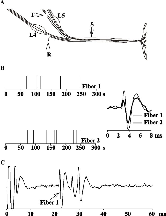
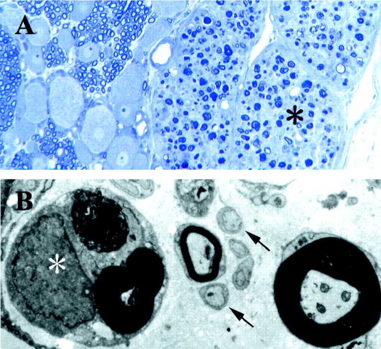
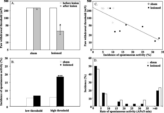
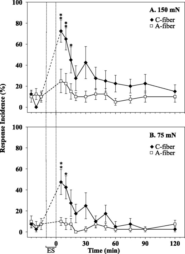
We demonstrated recently that uninjured C-fiber nociceptors in the L4 spinal nerve develop spontaneous activity after transection of the L5 spinal nerve. We postulated that Wallerian degeneration leads to an alteration in the properties of the neighboring, uninjured afferents from adjacent spinal nerves. To explore the role of degeneration of myelinated versus unmyelinated fibers, we investigated the effects of an L5 ventral rhizotomy in rat. This lesion leads to degeneration predominantly in myelinated fibers. Mechanical paw-withdrawal thresholds were assessed with von Frey hairs, and teased-fiber techniques were used to record from single C-fiber afferents in the L4 spinal nerve. Behavioral and electrophysiological data were collected in a blinded manner. Seven days after surgery, a marked decrease in withdrawal thresholds was observed after the ventral rhizotomy but not after the sham operation. Single fiber recordings revealed low-frequency spontaneous activity in 25% of the C-fiber afferents 8-10 d after the lesion compared with only 11% after sham operation. Paw-withdrawal thresholds were inversely correlated with the incidence of spontaneous activity in high-threshold C-fiber afferents. In normal animals, low-frequency electrocutaneous stimulation at C-fiber, but not A-fiber, strength produced behavioral signs of secondary mechanical hyperalgesia on the paw. These results suggest that degeneration in myelinated efferent fibers is sufficient to induce spontaneous activity in C-fiber afferents and behavioral signs of mechanical hyperalgesia. Ectopic spontaneous activity from injured afferents was not required for the development of the neuropathic pain behavior. These results provide additional evidence for a role of Wallerian degeneration in neuropathic pain.
Figures
Similar articles
-
Early onset of spontaneous activity in uninjured C-fiber nociceptors after injury to neighboring nerve fibers.J Neurosci. 2001 Apr 15;21(8):RC140. doi: 10.1523/JNEUROSCI.21-08-j0002.2001. J Neurosci. 2001. PMID: 11306646 Free PMC article.
-
Mechanical hyperalgesia after an L5 spinal nerve lesion in the rat is not dependent on input from injured nerve fibers.Pain. 2000 Apr;85(3):493-502. doi: 10.1016/S0304-3959(00)00250-5. Pain. 2000. PMID: 10781924
-
Mechanical hyperalgesia after an L5 ventral rhizotomy or an L5 ganglionectomy in the rat.Pain. 2002 Mar;96(1-2):63-72. doi: 10.1016/s0304-3959(01)00429-8. Pain. 2002. PMID: 11932062
-
A role for uninjured afferents in neuropathic pain.Sheng Li Xue Bao. 2008 Oct 25;60(5):605-9. Sheng Li Xue Bao. 2008. PMID: 18958368 Review.
-
Electrophysiological characterization of ectopic spontaneous discharge in axotomized and intact fibers upon nerve transection: a role in spontaneous pain?Pflugers Arch. 2022 Apr;474(4):387-396. doi: 10.1007/s00424-021-02655-7. Epub 2022 Jan 28. Pflugers Arch. 2022. PMID: 35088129 Review.
Cited by
-
Clinical indicators to identify neuropathic pain in low back related leg pain: a modified Delphi study.BMC Musculoskelet Disord. 2020 Sep 8;21(1):601. doi: 10.1186/s12891-020-03600-y. BMC Musculoskelet Disord. 2020. PMID: 32900367 Free PMC article.
-
An approach to identify microRNAs involved in neuropathic pain following a peripheral nerve injury.Front Neurosci. 2014 Aug 29;8:266. doi: 10.3389/fnins.2014.00266. eCollection 2014. Front Neurosci. 2014. PMID: 25221468 Free PMC article.
-
Fabry disease Schwann cells release p11 to induce sensory neuron hyperactivity.JCI Insight. 2024 Mar 7;9(8):e172869. doi: 10.1172/jci.insight.172869. JCI Insight. 2024. PMID: 38646936 Free PMC article.
-
Fight fire with fire: Neurobiology of capsaicin-induced analgesia for chronic pain.Pharmacol Ther. 2021 Apr;220:107743. doi: 10.1016/j.pharmthera.2020.107743. Epub 2020 Nov 10. Pharmacol Ther. 2021. PMID: 33181192 Free PMC article. Review.
-
Plasticity in intact A delta- and C-fibers contributes to cold hypersensitivity in neuropathic rats.Neuroscience. 2007 Nov 30;150(1):182-93. doi: 10.1016/j.neuroscience.2007.09.002. Epub 2007 Sep 19. Neuroscience. 2007. PMID: 17945425 Free PMC article.
References
-
- Ali Z, Ringkamp M, Hartke TV, Chien HF, Flavahan NA, Campbell JN, Meyer RA. Uninjured C-fiber nociceptors develop spontaneous activity and alpha adrenergic sensitivity following L6 spinal nerve ligation in the monkey. J Neurophysiol. 1999;81:455–466. - PubMed
-
- Ali Z, Meyer RA, Belzberg AJ. Neuropathic pain after C7 spinal nerve transection in man. Pain. 2002;96:41–47. - PubMed
-
- Bennett GJ, Xie Y-K. A peripheral mononeuropathy in rat that produces disorders of pain sensation like those seen in man. Pain. 1988;33:87–107. - PubMed
-
- Black JA, Cummins TR, Plumpton C, Chen YH, Hormuzdiar W, Clare JJ, Waxman SG. Upregulation of a silent sodium channel after peripheral, but not central, nerve injury in DRG neurons. J Neurophysiol. 1999;82:2776–2785. - PubMed
-
- Blumberg H, Jänig W. Discharge pattern of afferent fibers from a neuroma. Pain. 1984;20:335–353. - PubMed
Publication types
MeSH terms
Grants and funding
LinkOut - more resources
Full Text Sources