Review
. 2002 Oct;71(4):695-714.
doi: 10.1086/342730. Epub 2002 Aug 26.

Neocentromeres: role in human disease, evolution, and centromere study

Affiliations
Review

Neocentromeres: role in human disease, evolution, and centromere study

David J Amor et al. Am J Hum Genet. 2002 Oct.

Abstract

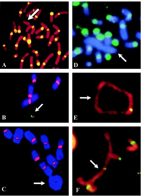
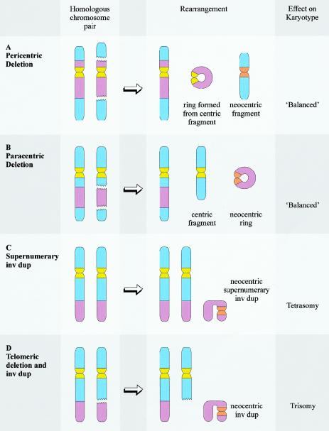
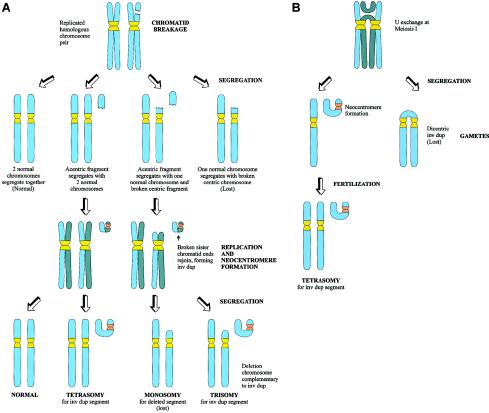
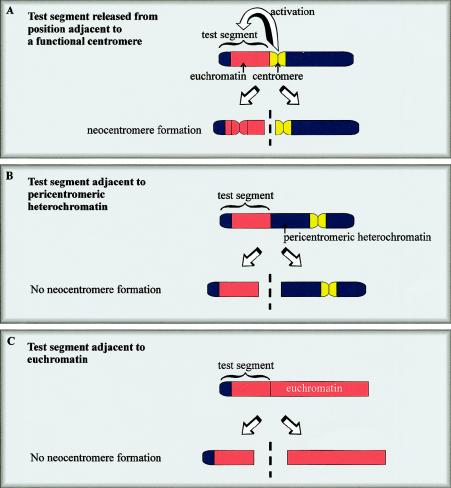
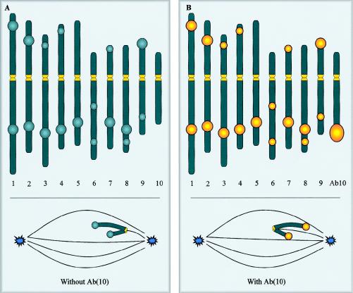
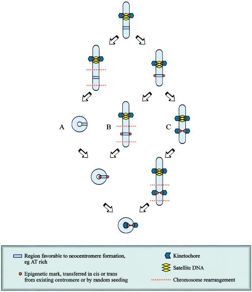
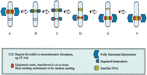
The centromere is essential for the proper segregation and inheritance of genetic information. Neocentromeres are ectopic centromeres that originate occasionally from noncentromeric regions of chromosomes. Despite the complete absence of normal centromeric alpha-satellite DNA, human neocentromeres are able to form a primary constriction and assemble a functional kinetochore. Since the discovery and characterization of the first case of a human neocentromere in our laboratory a decade ago, 60 examples of constitutional human neocentromeres distributed widely across the genome have been described. Typically, these are located on marker chromosomes that have been detected in children with developmental delay or congenital abnormalities. Neocentromeres have also been detected in at least two types of human cancer and have been experimentally induced in Drosophila. Current evidence from human and fly studies indicates that neocentromere activity is acquired epigenetically rather than by any alteration to the DNA sequence. Since human neocentromere formation is generally detrimental to the individual, its biological value must lie beyond the individual level, such as in karyotype evolution and speciation.


Figures

Figure  1
Figure 1
FISH analysis of human neocentric chromosomes. A, Patient cells with a 10q25 neocentromere-containing mardel(10) chromosome (arrow), using pancentromeric α-satellite probe, demonstrating absence of α-satellite (yellow) on the marker chromosome. Image taken from Voullaire et al. (1993). B, A stable, <2-Mb HAC (arrow) engineered from the mardel(10) chromosome shown in A (Saffery et al. 2001). Chromosome staining is with DAPI. Image courtesy of L. Wong. C–F, Partial metaphases of well-differentiated liposarcoma cases, using FISH with a pancentromeric α-satellite probe (C and D) and immunostaining with anticentromere antibody (E and F). Arrows indicate the supernumerary rings and large rod marker chromosomes. FISH signals (red in C or green in D) with the α-satellite probe are observed on all chromosomes except the supernumerary ring (C) and large marker (D). Positive staining with the anticentromere antibody (yellow or green) is observed on all chromosomes including the supernumerary analphoid ring (E) and large marker (F). Images courtesy of F. Pedeutour and N. Sirvent.
Figure  2
Figure 2
Sites of formation of constitutional neocentromeres within the human genome. A total of 60 cases, originating from 16 different human chromosomes, have been described. The mapped positions of each of the neocentromere cases are indicated by bars to the right of the chromosome ideograms. Longer bars indicate neocentromere sites that have not been precisely localized. Hatch marks on chromosomes 1, 9, and Y represent blocks of constitutive heterochromatin. Adapted from Choo (2001a).
Figure  3
Figure 3
Chromosome rearrangements commonly associated with neocentromere formation. Neocentromere formation is typically associated with a chromosome rearrangement resulting in the generation of a fragment that lacks a conventional centromere. The most common chromosome rearrangements are interstitial deletions (A and B) and inverted duplications (C and D). Interstitial deletions are typically associated with the formation of a ring chromosome, which may be necessary to stabilize the broken chromosome ends. The ring chromosome may be derived from the centric deletion chromosome (in the case of pericentric deletions, shown in A) or from the neocentric fragment (in the case of paracentric deletions, shown in B). When neocentromere formation results from an interstitial deletion, the resulting karyotype is usually “balanced” at a cytogenetic level. However, phenotypic effect may result from a “ring syndrome,” leading to mosaicism for one of the fragments, or from interruption of critical genes at the sites of chromosome breakage or neocentromere formation. Inverted duplications can be supernumerary to the original karyotype (C) (resulting in tetrasomy for the duplicated segment) or accompanied by a complementary deletion (D) (resulting in trisomy for the duplicated segment).
Figure  4
Figure 4
Possible mechanisms of formation of neocentric inverted duplication (“inv dup”) marker chromosomes. A, Formation at mitosis. Chromatid breakage is followed by segregation of the acentric fragment and the centric fragment (or deletion chromosome). Following replication, the broken sister chromosome ends of the acentric fragment join to form a mirror image inv dup chromosome, with neocentromere formation occurring on one of the two arms of the inv dup. The centric fragment will usually be lost because of instability; however, stabilization of the broken end of the fragment will occasionally allow the fragment to survive. Segregation of the inv dup with the centric fragment will lead to trisomy for the chromosome segment involved, whereas, if the inv dup segregates with two normal chromosome homologues, tetrasomy will result. B, Formation at meiosis. Formation of an acentric inv dup fragment occurs because of anomalous crossing-over during meiosis I. After segregation, the dicentric fragment will be lost, but the acentric fragment may be “rescued” by neocentromere formation. After fertilization, zygotes containing the inv dup will be tetrasomic for the segment involved in the inv dup.
Figure  5
Figure 5
Generation of neocentromeres in Drosophila. A test segment comprising telomeric heterochromatin and euchromatin forms a functional neocentromere when released from a site immediately adjacent to a normal centromere (A). One model suggests that centromere activity or “centromere imprinting factor” spreads from the existing centromere to the neighboring test DNA, where it activates or imparts a stable centromeric state that can come into independent existence when this DNA is subsequently released. When the same fragment is released from sites adjacent to pericentromeric heterochromatin (B) or euchromatin (C), a neocentromere does not form.
Figure  6
Figure 6
Maize knob neocentromeres and the maize karyotype, indicating some of the more common sites of heterochromatic knobs on the 10 chromosomes (Rhoades 1950). In the presence of a normal chromosome 10 (A), the knobs are inactive and lag behind the normal centromere at meiosis (inset). When the normal chromosome 10 is replaced by Ab(10), the knobs become neocentromeres. These neocentromeres bind the spindle microtubules in a lateral rather than an end-on manner and migrate towards the spindle pole in advance of the normal centromeres that remain active (B). Yellow indicates an active centromere or neocentromere.
Figure  7
Figure 7
Formation of human neocentromeres. Human neocentromeres can form in either meiosis or mitosis, by a mechanism that probably involves the acquisition of a centromere-specific epigenetic mark, followed by formation of a functional kinetochore. Certain chromosomal regions are predisposed to neocentromere formation, possibly because of AT content, heterochromatic qualities, or “centromere-correct” replication timing. The formation of a marker chromosome containing a neocentromere is dependent on three steps: (1) rearrangement of the chromosome, generating an acentric fragment (in this example, rearrangement is a paracentric deletion resulting in the formation of a centric deletion chromosome and a neocentric ring); (2) acquisition of the epigenetic mark required for centromere determination; and (3) formation of a functional kinetochores. The timing of these events in relation to each other is unclear. It is possible that chromosome rearrangement is the initial event (path A), followed by acquisition of the epigenetic mark and formation of the kinetochore. Alternatively, the chromosome arrangement may occur between acquisition of the epigenetic mark and formation of the kinetochore (path B), or, less likely, the chromosome rearrangement may be consequent to the formation of a functional kinetochore (path C).
Figure  8
Figure 8
Evolutionary repositioning of centromeres by neocentromere formation. The initial event in centromere repositioning may be an impairment of function of the original centromere (B), possibly leading to a reduction of lateral inhibition. A neocentromere may then form via epigenetic mechanisms not involving alteration to the primary DNA sequence at a favorable site (C). The initial neocentromere may be imperfect, but, in subsequent generations, selection pressure improves kinetochore maturation through duplication of existing sequence or accumulation of repetitive DNA from other sources (D–F). The original satellited centromeric DNA would subsequently contract in the absence of selection pressure and ultimately disappear (D–F).

References

    1. Aagaard L, Schmid M, Warburton P, Jenuwein T (2000) Mitotic phosphorylation of SUV39H1, a novel component of active centromeres, coincides with transient accumulation at mammalian centromeres. J Cell Sci 113:817–829 - PubMed
    1. Abeliovich D, Yehuda O, Ben-Neriah S, Kapelushnik Y, Ben-Yehuda D (1996) dup(10q) lacking alpha-satellite DNA in bone marrow cells of a patient with acute myeloid leukemia. Cancer Genet Cytogenet 89:1–6 - PubMed
    1. Ananiev EV, Phillips RL, Rines HW (1998) A knob-associated tandem repeat in maize capable of forming fold-back DNA segments: are chromosome knobs megatransposons? Proc Natl Acad Sci USA 95:10785–10790 - PMC - PubMed
    1. Assumpção JG, Berkofsky-Fessler W, Campos NV, Macial-Guerra AT, Li S, Melaragno MI, de Mello MP, Warburton PE. Identification of a neocentromere in a rearranged Y chromosome with no detectable DY23 centromeric sequence. Am J Med Genet (in press) - PubMed
    1. Barbi G, Kennerknecht I, Wohr G, Avramopoulos D, Karadima G, Petersen MB (2000) Mirror-symmetric duplicated chromosome 21q with minor proximal deletion, and with neocentromere in a child without the classical Down syndrome phenotype. Am J Med Genet 91:116–122 - PubMed

Publication types

LinkOut - more resources