Review
. 2002 Aug;90(2):157-67.
doi: 10.1093/aob/mcf180.

Biophysical limitation of cell elongation in cereal leaves

Affiliations
Review

Biophysical limitation of cell elongation in cereal leaves

Wieland Fricke. Ann Bot. 2002 Aug.

Abstract

Grass leaves grow from the base. Unlike those of dicotyledonous plants, cells of grass leaves expand enclosed by sheaths of older leaves, where there is little or no transpiration, and go through developmental stages in a strictly linear arrangement. The environmental or developmental factor that limits leaf cell expansion must do so through biophysical means at the cellular level: wall-yielding, water uptake and solute supply are all candidates. This Botanical Briefing looks at the possibility that tissue hydraulic conductance limits cell expansion and leaf growth. A model is presented that relates pathways of water movement in the elongation zone of grass leaves to driving forces for water movement and to anatomical features. The bundle sheath is considered as a crucial control point. The relative importance of these pathways for the regulation of leaf growth and for the partitioning of water between expansion and transpiration is discussed.


Figures
None
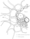
Fig. 1. Elongating cells embedded in a tissue can be located considerable distances and many cell layers away from the nearest water source. The diagram shows a cross‐sectional view of the area next to a major vascular bundle in the leaf elongation zone of a grass, barley. Water and solutes exiting the protoxylem (PX) or metaxylem (MX) have to pass several layers of cells before reaching peripherally located tissue (here, mesophyll, MS). Epidermal cells are located even further away (compare Figs 3 and 4). During its passage, water has to cross two bundle sheaths, the mestome sheath (MSH) and the parenchymatous bundle sheath (PBS). The walls of the mestome sheath may be suberized, as indicated by the bold line. Water can move along an apoplastic path or along a combined symplastic/transcellular path. For simplicity, each path is shown only for one direction of water flow.
None
Fig. 2. Profile of relative elemental growth rate (REGR) along the growth zone of leaf three of barley. Plants were grown under control conditions, subjected to 120 mm NaCl (Fricke and Peters, 2002) or grown under source‐reduced conditions (Fricke, 2002). The latter was achieved by removing the blades of older leaves at the time leaf three emerged from encircling sheaths. Profiles of REGR were determined by pin‐pricking and corrected for the reduction in leaf elongation velocity due to pricking.
None
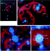
Fig. 3. Cross‐sectional view of the third leaf of barley at 10–12 mm (A and B) and 26–30 mm (C) from the point of leaf insertion, and halfway along the emerged part of the blade (D). At 26–30 mm from the point of leaf insertion, cells expand at near maximum relative rates, and growth‐associated water potential gradients are largest. The photographs show autofluorescence of lignified xylem vessels or vein tissue (blue) and mesophyll (red). Pictures were taken with an Axioscope (Zeiss, Germany: excitation filter, G 365; chromatic beam splitter, FT 395; barrier filter, LP 420). Note, the change in autofluorescence of metaxylem vessels between B/C and D. Cross‐sections of the entire leaf (A) and of larger leaf sections (B and C) show that not every vein and surrounding tissue is supplied with water by its ‘own’ vessels. Instead, almost 100 % of water is supplied by six large lateral veins (LV) and the midrib (MR). Along the elongation zone, water is conducted within protoxylem vessels (PX); the larger metaxylem vessels (MX) are lignified and thought to be fully functional only beyond the elongation zone (D). Protoxylem vessels are often separated by several cell layers from the mestome sheath (MSH) and parenchymatous bundle sheath (PBS). In contrast, metaxylem vessels border always directly at the mestome sheath. PH, Phloem; ABEP, abaxial epidermis; ADEP, adaxial epidermis. Bar as shown in part A represents 200 µm (A), 50 µm (B and C) or 25 µm (D).
None
Fig. 4. Cross‐sectional view of the MR tissue of the mature blade of leaf three of barley, following staining with berberine hemisulfate (Brundrett et al., 1988). Leaves were viewed under fluorescence, with the same filter setting as in Fig. 3, and counter‐stained with aniline blue (A) or not counter‐stained (B). Following counter‐staining with aniline blue, berberine hemisulfate stains lignified walls bright yellow, Casparian bands intense yellow‐white and suberin blue white or blue (Brundrett et al., 1988). Note the bright fluorescence of metaxylem vessels, and the fluorescence of radial walls of the mestome sheath (MSH) and of the border region separating xylem and phloem. Fluorescence at the leaf surface points to guard cells or sclerenchymateous tissue. Red fluorescence in B originates from chlorophyll (mesophyll). Bars = 25 µm (A) and 50 µm (B). PBS, Parenchymatous bundle sheath.
None
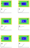
Fig. 5. Model for water transport between xylem and peripheral cells in the elongation zone of grass leaves. Various scenarios are considered (A–F). Tissue (cross‐sectional view) is divided into tissue located inside vascular bundles (blue; xylem, phloem, parenchyma), bundle sheaths (purple; mestome sheath and parenchymatous bundle sheath), mesophyll (green) and epidermis (yellow). Water moves along a combined transcellular/symplastic path, i.e. crossing protoplasts (black arrows; the length of arrows in A–F indicates whether a particular pathway applies only for passage of the bundle sheaths or for the entire path between vein tissue and epidermis), or along a predominantly apoplastic path (white arrows). Proposed gradients in water potential (ψ) are also shown. A, Water reaches peripheral cells along a symplastic/transcellular path, and a large drop in water potential occurs at the bundle sheath (lowest hydraulic conductance), due to suberization of the mestome sheath and a low hydraulic conductivity (aquaporin regulation) of cells. For the same reason, water moving towards the xylem (transpiration tension) has to cross the bundle sheath along the symplastic/transcellular path. Growth‐associated Δψ does not translate into apoplast tension, hence there is no outward‐directed hydraulic force. B, Same pathways of water movement as in A, except that layers of small parenchymatous cells, which are located within the bundle, represent the main hydraulic barrier (see drop in ψ; compare Tang and Boyer, 2002). C, As in A, except that water moves peripherally along the symplastic/transcellular path only at the bundle sheath region (as indicated by short black arrow) due to apoplastic barriers. Before and beyond the bundle sheath, water moves apoplastically (for simplicity not shown), either driven hydraulically by growth‐induced apoplast tension (outward) or by xylem tension (inward). D, This is the only scenario with insignificant growth‐associated Δψ. This could be due to overestimation of osmotic forces based on in vitro analyses of osmolality—if cells have reflection coefficients for main osmolytes that are <<1·0—or due to consistent underestimation of turgor pressure in plants prepared for turgor analyses (see Table 2) regardless of residual elongation velocity. Water moves in either direction mainly along the low‐resistance apoplast path, and the bundle sheath represents neither an apoplastic nor an osmotic barrier for water movement. E, As in A, except that the bundle sheath does not represent an apoplastic barrier for water movement. Despite this, water moves peripherally along the symplastic/transcellular path since growth‐associated ψ does not cause apoplast tension (but apoplast solute potential which cannot drive water movement). Water potential drops at the bundle sheath due to low conductivity of cells, most likely because of low aquaporin activity (this is also implicit in A). Inward flow of water is driven by transpiration tension. This scenario implies that during periods of positive xylem pressure (no transpiration) water can move peripherally along the apoplast and that ψ gradients may be insignificant (not indicated in figure). F, As in E, except that low‐conducting cells in the xylem parenchyma represent the major hydraulic constriction.

Similar articles

Cited by

References

    1. AcevedoE, Hsiao TC, Henderson DW.1971. Immediate and subsequent growth responses of maize leaves to changes in water status. Plant Physiology 48: 631–636. - PMC - PubMed
    1. ArifH, Tomos AD.1993. Control of wheat leaf growth under saline conditions. In: Lieth H, Al Masoom H, eds. Towards the rational use of high salinity tolerant plants. Vol 2 Dordrecht, The Netherlands: Kluwer Academic Publishers, 45–52.
    1. BarlowEWR.1986. Water relations of expanding leaves. Australian Journal of Plant Physiology 13: 45–58.
    1. Ben‐Haj‐SalahH, Tardieu F.1995. Temperature affects expansion rate of maize leaves without changes in spatial distribution of cell length. Plant Physiology 109: 861–870. - PMC - PubMed
    1. BernsteinN, Läuchli A, Silk WK.1993. Kinematics and dynamics of sorghum (Sorghum bicolor L.) leaf development at various Na/Ca salinities. I. Elongation growth. Plant Physiology 103: 1107–1114. - PMC - PubMed

Publication types

MeSH terms