

Immunosuppressive properties of human amniotic membrane for mixed lymphocyte reaction
- PMID: 12197887
- PMCID: PMC1906465
- DOI: 10.1046/j.1365-2249.2002.01945.x
Immunosuppressive properties of human amniotic membrane for mixed lymphocyte reaction
Abstract
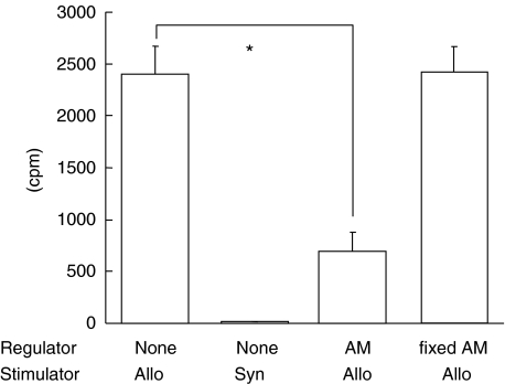
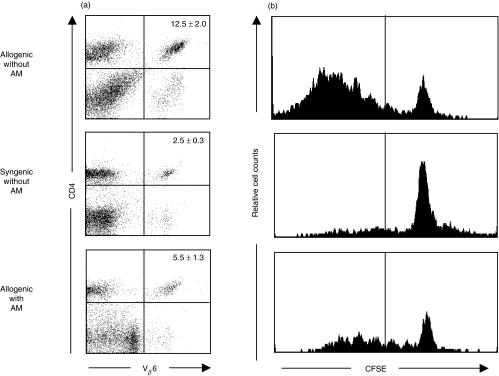
The combination of allograft limbal transplantation (ALT) and amniotic membrane transplantation (AMT) has been applied in the treatment of severe ocular surface diseases. The beneficial effect of this combination has been thought to result from possible immunosuppressive ability of amniotic membrane (AM). However, the mechanisms of any such ability remain unknown. In this study, we investigated whether human AM has the ability to suppress allo-reactive T cell responses in vitro. For mixed lymphocyte reaction (MLR), lymphocytes isolated from lymph nodes of C57BL/6 mice (Mls1b, Vbeta6+) were cultured with irradiated splenocytes from DBA/2 mice (Mls1a, Vbeta6-) with or without human AM. For carboxyfluorescein diacetate succinimidyl ester (CFSE) experiments, responder lymph node cells were labelled with a stable intracellular fluorescent dye and cultured with irradiated stimulator cells. The ratio of responder Vbeta6+ T cells was then determined by FACS analysis, and the division profiles of responder Vbeta6+ T cells were analysed by CFSE content. Furthermore, Th1 and Th2 cytokine synthesis by allo-reactive T cells in MLR culture supernatants was determined by enzyme-linked immunosorbent assay (ELISA). Addition of AM to the MLR culture resulted in the significant inhibition of thymidine incorporation compared with control culture lacking AM. The population of responder CD4+Vbeta6+ T cells was significantly reduced in the AM-treated culture in comparison to control. CFSE analysis revealed less division and lower proliferation of responder CD4+Vbeta6+ T cells in cultures with AM than without. In addition, allo-rective T cell synthesis of both Th1 (IL-2 and IFNgamma) and Th2 (IL-6 and IL-10) type cytokine was significantly decreased in the presence of AM. These results indicate that human AM has the ability to suppress allo-reactive T cells in vitro. This inhibitory effect likely contributes to the success of the ALT-AMT combination.
Figures
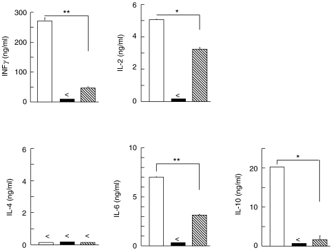
 ), allogenic MLR without AM (□) and syngenic MLR without AM (▪) were examined by murine Th1 and Th2 cytokine-specific ELISA. Following 2 day incubation, IL-2 synthesis level was measured. In case of IFNγ, IL-6 and IL-10, supernatants harvested from 4 day cultures were subjected to cytokine-specific ELISA. Data represent mean ± SEM from 1 experiment with duplicated wells. (<not detectable; *P < 0·01; **P < 0·001)
References
-
- Kinoshita S, Kiorpes T, Friend J, et al. Limbal epithelium in ocular surface wound healing. Invest Ophthalmol Vis Sci. 1982;23:73–80. - PubMed
-
- Shapiro M, Friend J, Thoft R. Corneal re-epithelialization from the conjunctiva. Invest Ophthalmol Vis Sci. 1981;21:135–42. - PubMed
-
- Cotsarelis G, Cheng S, Dong G, et al. Existence of slow-cycling limbal epithelial basal cells that can be preferentially stimulated to proliferate: implications on epithelial stem cells. Cell. 1989;57:201–9. - PubMed
-
- Huang A, Tseng S. Corneal epithelial wound healing in the absence of limbal epithelium. Invest Ophthalmol Vis Sci. 1991;32:96–105. - PubMed
MeSH terms
Substances
LinkOut - more resources
Full Text Sources
Other Literature Sources
Research Materials
