. 2002 Sep 2;21(17):4411-9.
doi: 10.1093/emboj/cdf445.

A mitochondrial specific stress response in mammalian cells

Affiliations

A mitochondrial specific stress response in mammalian cells

Quan Zhao et al. EMBO J. .

Abstract

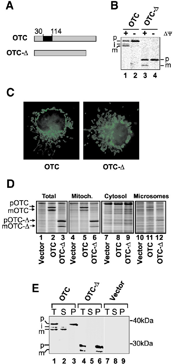
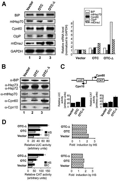
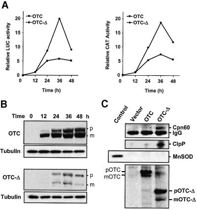
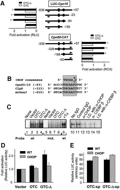
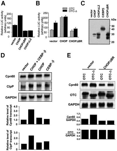
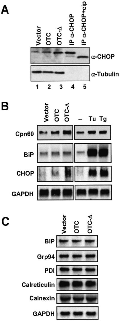
Cells respond to a wide variety of stresses through the transcriptional activation of genes that harbour stress elements within their promoters. While many of these elements are shared by genes encoding proteins representative of all subcellular compartments, cells can also respond to stresses that are specific to individual organelles, such as the endoplasmic reticulum un folded protein response. Here we report on the discovery and characterization of a mitochondrial stress response in mammalian cells. We find that the accumulation of unfolded protein within the mitochondrial matrix results in the transcriptional upregulation of nuclear genes encoding mitochondrial stress proteins such as chaperonin 60, chaperonin 10, mtDnaJ and ClpP, but not those encoding stress proteins of the endoplasmic reticulum. Analysis of the chaperonin 60/10 bidirectional promoter identified a CHOP element as the mitochondrial stress response element. Dominant-negative mutant forms of CHOP and overexpression of CHOP revealed that this transcription factor, in association with C/EBPbeta, regulates expression of mitochondrial stress genes in response to the accumulation of unfolded proteins.


Figures

None
Fig. 1. Establishment of a mitochondrial UPR. (A) Diagrammatic representation of the wild-type OTC and deletion mutant (OTC-Δ) from which the carbamyl phosphate-binding domain (amino acids 30–114 of mature OTC) has been removed. (B) OTC and OTC-Δ are imported into mitochondria in vivo. COS-7 cells transfected with OTC (lanes 1 and 2) or OTC-Δ (lanes 3 and 4) were labelled with [35S]methionine 36 h after transfection in the presence or absence of rhodamine 6G to disrupt the mitochondrial inner membrane potential (Δψ). Cell extracts were immunoprecipitated with antibodies specific for OTC and subjected to SDS–PAGE and PhosphorImager analysis (p, precursor; i, processing intermediate; m, mature). (C) OTC-Δ localizes to mitochondria. Immunofluorescence microscopy of cells transfected with OTC or OTC-Δ was employed using antibodies against OTC. (D) OTC and OTC-Δ are associated with the mitochondrial fraction in radiolabelled cells. Cells were transfected with vector alone or vector expressing OTC or OTC-Δ and, 32 h later, radiolabelled for 4 h. Cells were harvested and subjected to subcellular fractionation, SDS–PAGE and PhosphorImager analysis. Radiolabelled forms of precursor OTC/OTC-Δ and matrix-processed OTC/OTC-Δ are indicated. (E) OTC-Δ forms aggregates in mitochondria. Cells expressing OTC or OTC-Δ were harvested 36 h post-transfection. The total mitochondrial fraction (T) was separated into 0.5% (w/v) Triton X-100-soluble (S) and -insoluble (P) fractions, and subjected to SDS–PAGE followed by western blot analysis using OTC antibodies.
None
Fig. 2. Transcriptional activation of genes by unfolded proteins in mitochondria. (A) Northern blot analysis of total RNA isolated from COS-7 cells transfected with vector OTC or OTC-Δ constructs (left panel). The blot was hybridized sequentially with radiolabelled Cpn60, Cpn10, ClpP, BiP, mtHsp70, mtDnaJ and control GAPDH cDNA probes. The change in transcript levels normalized against GAPDH levels was quantitated relative to that found in cells transfected with vector alone (right panel). (B) Increase in the levels of chaperones within mitochondria. Immunoblot analysis of various stress proteins from equal amounts of whole-cell lysates from COS-7 cells transfected with vector, OTC or OTC-Δ. The blot was sequentially probed with antibodies against Cpn60, Cpn10, mtHsp70, Hsp72/73 and BiP. (C) Activation of the Cpn60/10 bidirectional promoter by mitochondrial stress. A reporter construct was used whereby the Cpn10 side of the bidirectional promoter regulates LUC expression and the Cpn60 side regulates CAT expression (top panel). COS-7 cells co-transfected with vector, OTC or OTC-Δ, Cpn60/10 reporter plasmid and β-galactosidase plasmid were assayed 36 h after transfection for LUC and CAT activities and normalized against β-galactosidase activity. Data represent the mean ± SEM from experiments performed in triplicate. (D) Activation of Cpn60/10 promoter by heat shock and mitochondrial stress is additive. COS-7 cells were co-transfected with vector, OTC or OTC-Δ, the Cpn60/10 reporter construct, and β-galactosidase plasmid. After 12 h, cells were split into two plates and 6 h later one set was heat shocked (HS, black bars) at 43°C for 1 h and the others left at 37°C (C, grey bars). Cell extracts were assayed after 16 h for LUC and CAT activities relative to β-galactosidase activity (left side). The fold activation due to HS compared with controls of the Cpn60/10 promoter in either orientation (Cpn60/CAT or Cpn10/LUC) is shown (right side).
None
Fig. 3. MSR is transient and correlates with the level of aggregated protein in mitochondria. (ACpn60 and Cpn10 promoter activation is transient. COS-7 cells were co-transfected with the Cpn60/10 promoter construct along with constructs expressing OTC or OTC-Δ and β-galactosidase plasmid. Cells were harvested at the indicated times and CAT and LUC activity relative to β-galactosidase activity was measured. (B) Changes in the steady-state levels of wild-type OTC and mutant OTC after transfection of cells. Equal amounts of protein from whole-cell extracts were subjected to SDS–PAGE, blotted and probed with an anti-OTC antibody, and anti-tubulin antibodies as a loading control. p, precursor of OTC; m, mature form of OTC. (C) Cpn60 and ClpP associate with mutant OTC-Δ. Mitochondrial proteins from COS-7 cells transfected with vector or OTC constructs were translabelled with [35S]methionine, immunoprecipitated with OTC antibodies and subjected to western blot analysis. The blot was probed with Cpn60 and ClpP antibodies (upper panels) and MnSOD antibodies as a control (middle panel; control on left side). The immunoprecipitated 35S- labelled wild-type OTC and OTC-Δ were visualized by PhosphorImager analysis of the filter (bottom panel).
None
Fig. 4. Identification of the MSR element in the Cpn60/10 promoter. (A) Promoter deletion analysis was carried out by making deletions from either the Cpn60 side (top panels) or Cpn10 side (bottom panels) of the Cpn60/10 bidirectional promoter. Deletions are shown as distances (bp) from the Cpn60 transcription start site. Cpn60 and Cpn10 transcription start sites are indicated by arrowheads. The relative position of the HSE is indicated by rectangles while circles indicate the CHOP element. The fold activation of the promoter construct in cells expressing OTC-Δ (solid bars) compared with those expressing wild-type OTC (open bars) is shown as relative luciferase (RLU) or CAT (RCA) activity. Data represents the mean ± SEM from experiments performed in triplicate. (B) Nucleotide sequence alignment of the CHOP consensus element in the Cpn60/10 and the putative ClpP and mtDnaJ promoters. Bold letters show identical nucleotides and numbers refer to distance from transcription initiation site of Cpn60 or the first base of the initiation codon for ClpP and mtDnaJ. The CHOP consensus binding site is shaded. (C) EMSA of nuclear extracts from COS-7 cells transfected with OTC constructs. A probe corresponding to a 30 bp Cpn60/10 promoter fragment that contains the CHOP-binding element (wt) and a mutant probe (mut.) containing an altered CHOP site were incubated with nuclear extracts isolated from cells transfected with various constructs or incubated with tunicamycin (Tu) as indicated and subjected to PAGE and PhosphorImager analysis. Immunocompetition and immunodepletion assays were performed using nuclear extracts of COS-7 cells transfected with OTC-Δ. Nuclear extracts were incubated with no IgG (lane 10), control (anti-β-galactosidase) IgG (lane 11), or with anti-CHOP or C/EBPβ antibodies (lanes 12 and 14), before adding radiolabelled wild-type probe. In addition, nuclear extracts were first immunodepleted (I.D.) of CHOP or C/EBPβ using antibodies attached to protein A–Sepharose followed by incubation with radiolabelled wild-type probe (lanes 13 and 15). (D) The CHOP site in the Cpn60/10 promoter is involved in the MSR response. The effect of introduction of a mutated CHOP element (CHOP) on the Cpn60/10 promoter activity was determined by co-transfecting cells with OTC constructs (vector, OTC, OTC-Δ) and wild-type (WT, black bars) or mutant (CHOP, grey bars) Cpn60/10 promoter constructs. Promoter activity was measured 36 h after transfection and is based on measurement of LUC expression (mean ± SEM). (E) The effect of expressing wild-type (OTC-sp) or mutant (OTC-Δ-sp) OTC lacking the mitochondrial signal peptide (sp) was measured by co-transfecting cells with OTC constructs and with wild-type (WT, black bars) or mutant (CHOP, grey bars) Cpn60/10 promoter reporter constructs. Promoter activity was measured 36 h after transfection and is based on measurement of LUC expression (mean ± SEM).
None
Fig. 5. Effect of CHOP expression on Cpn60/10 promoter activity. (A) Overexpression of CHOP and C/EBPβ activates the Cpn60/10 promoter. COS-7 cells were co-transfected with the Cpn60/10 promoter reporter construct and cDNA encoding C/EBPβ, as well as either vector alone, wild-type CHOP cDNA or CHOP constructs lacking either the basic DNA-binding region (CHOPΔBR) or the leucine zipper region (CHOPΔLZ). Relative LUC activity was measured 36 h after transfection. Results represent the mean of transfections performed in triplicate ± SEM. (B) CHOP-dependent induction of MSR. The effect of a dominant-negative mutant of CHOP (CHOPΔBR) on the MSR was measured by co-transfecting cells with the Cpn60/10 promoter reporter construct, OTC constructs (OTC, grey bars; OTC-Δ, black bars), cDNA encoding C/EBPβ and either vector alone, wild-type CHOP or CHOPΔBR cDNA. Relative LUC activities were measured in cell extracts 36 h after transfection and represent the mean of transfections performed in triplicate ± SEM. (C) Level of expression of transcription vectors. The expression of CHOP constructs and C/EBPβ by transfected cells (30 µg of total protein/lane) is shown by western blot analysis using specific antibodies. (D) Overexpression of CHOP and C/EBPβ increases the level of Cpn60 and ClpP transcripts. COS-7 cells were transfected with cDNA encoding CHOP and C/EBPβ and total RNA extracted from cells subjected to northern blot analysis. The blot was sequentially hybridized with radiolabelled Cpn60, ClpP and GAPDH cDNA probes (top panels). Blots were quantitated relative to GAPDH by PhosphorImager analysis (lower panels) with the ratio of transcripts from cells transfected with vector alone being set at 1.0. (E) Induction of Cpn60 transcripts by MSR is ablated by a dominant-negative form of CHOP. The effect of transfection of COS-7 cells with vector alone or dominant-negative mutant CHOPΔBR on Cpn60 mRNA levels was measured by northern blot analysis from cells co-transfected with wild-type OTC and OTC-Δ. The levels of wild-type and mutant OTC transcripts were also determined as a control (upper panels). Bands were quantitated by PhosphorImager analysis relative to the levels of GAPDH (lower panels).
None
Fig. 6. CHOP induction and specificity of the MSR. (A) CHOP expression in response to mitochondrial stress. Extracts from cells transfected with vector, wild-type OTC or mutant OTC-Δ constructs were subjected to western blotting transfers and probed with antibodies against CHOP (α-CHOP) and tubulin (α-tubulin) as control. Extracts of cells transfected with wild-type CHOP cDNA were subjected to immunoprecipitation with antibodies against CHOP (IP α-CHOP) and half of the immunoprecipitate was treated with calf intestinal phosphatase (IP α-CHOP + cip) followed by the western blot analysis probed with antibodies against CHOP. (B) Specificity of MSR and the ER UPR. RNA was isolated from cells transfected with vector, wild-type OTC, OTC-Δ or with agents that trigger the ER stress response, tunicamycin (Tu) and thapsigargin (Tg) and subjected to northern blot analysis. The blot was sequentially hybridized with radiolabelled Cpn60, BiP, CHOP and GAPDH cDNA probes. (C) MSR does not affect expression of genes for ER stress proteins. RNA isolated from cells transfected with vector, wild-type OTC or OTC-Δ was subjected to northern blot analysis using probes corresponding to transcripts encoding ER proteins as well as GAPDH.

References

    1. Andrews N.C. and Faller,D.V. (1991) A rapid micropreparation technique for extraction of DNA-binding proteins from limiting numbers of mammalian cells. Nucleic Acids Res., 19, 2499. - PMC - PubMed
    1. Bartlett J.D., Luethy,J.D., Carlson,S.G., Sollott,S.J. and Holbrook,N.J. (1992) Calcium ionophore A23187 induces expression of the growth arrest and DNA damage inducible CCAAT/enhancer-binding protein (C/EBP)-related gene, gadd153. Ca2+ increases transcriptional activity and mRNA stability. J. Biol. Chem., 267, 20465–20470. - PubMed
    1. Biswas G., Adebanjo,O.A., Freedman,B.D., Anandatheerthavarada,H.K., Vijayasarathy,C., Zaidi,M., Kotlikoff,M. and Avadhani,N.G. (1999) Retrograde Ca2+ signaling in C2C12 skeletal myocytes in response to mitochondrial genetic and metabolic stress: a novel mode of inter-organelle crosstalk. EMBO J., 18, 522–533. - PMC - PubMed
    1. Bruhat A., Jousse,C., Wang,X.Z., Ron,D., Ferrara,M. and Fafournoux,P. (1997) Amino acid limitation induces expression of CHOP, a CCAAT/enhancer binding protein-related gene, at both tran scriptional and post-transcriptional levels. J. Biol. Chem., 272, 17588–17593. - PubMed
    1. Calfon M., Zeng,H., Urano,F., Till,J.H., Hubbard,S.R., Harding,H.P., Clark,S.G. and Ron,D. (2002) IRE1 couples endoplasmic reticulum load to secretory capacity by processing the XBP-1 mRNA. Nature, 415, 92–96. - PubMed

Publication types

MeSH terms