. 2002 Sep 2;21(17):4480-90.
doi: 10.1093/emboj/cdf472.

Vesicle transmembrane potential is required for translocation to the cytosol of externally added FGF-1

Affiliations

Vesicle transmembrane potential is required for translocation to the cytosol of externally added FGF-1

Jedrzej Małecki et al. EMBO J. .

Abstract

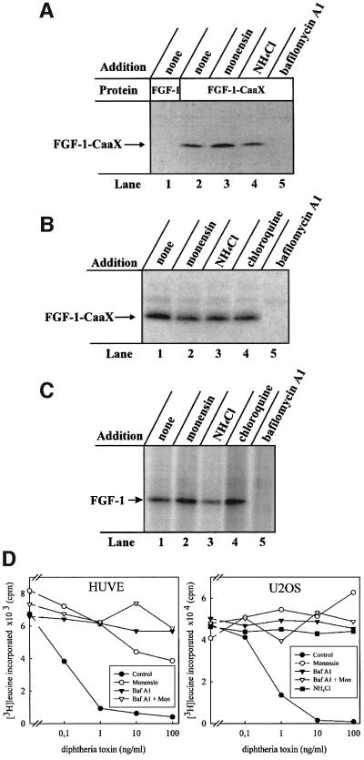
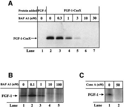
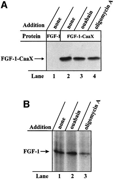
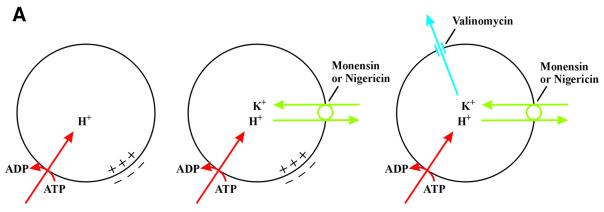
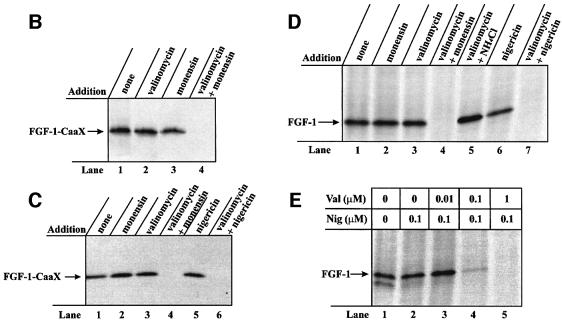
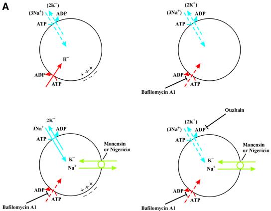
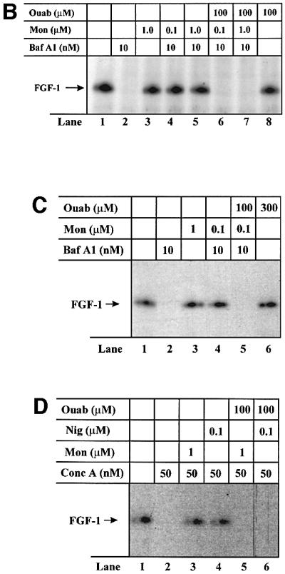
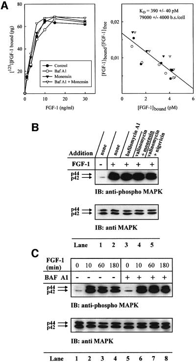
Externally added fibroblast growth factor-1 (FGF-1) is capable of crossing cellular membranes to reach the cytosol and the nucleus in a number of cell types. We have monitored the translocation of the growth factor by two methods: phosphorylation of FGF-1, and prenylation of an FGF-1 mutant that contains a C-terminal prenylation signal. Inhibition of endosomal acidification by ammonium chloride or monensin did not block the translocation of FGF-1, whereas bafilomycin A1, a specific inhibitor of vacuolar proton pumps, blocked translocation completely. A combination of ionophores expected to dissipate the vesicular membrane potential (valinomycin plus monensin) also fully inhibited the translocation. The inhibition of translocation by bafilomycin A1 was overcome in the presence of monensin or nigericin, while ouabain blocked translocation under these conditions. The data indicate that translocation of FGF-1 to cytosol occurs from the lumen of intracellular vesicles possessing vacuolar proton pumps, and that a vesicular membrane potential is required. Apparently, activation of vesicular Na+/K+-ATPase by monensin or nigericin generates a membrane potential that can support translocation when the proton pump is blocked.


Figures

None
Fig. 1. Effect of drugs that neutralize acidic cellular compartments on the ability of exogenous FGF-1 to become prenylated and phosphorylated in vivo. (A) Serum-starved CPAE cells were pre-incubated with radiolabelled mevalonic acid and lovastatin, and then incubated with 10 U/ml heparin and 100 ng/ml FGF-1 (lane 1) or 100 ng/ml FGF-1–CaaX (lanes 2–5) for 6 h in the absence (lanes 1 and 2) or presence of 1 µM monensin (lane 3), 20 mM NH4Cl (lane 4) or 10 nM bafilomycin A1 (lane 5). After lysis, the material adsorbed onto heparin–Sepharose was analysed by SDS–PAGE and fluorography. The arrow indicates the migration of prenylated FGF-1–CaaX. (B) NIH 3T3 cells were pre-treated as in (A). The incubation was carried out in the absence (lane 1) or presence of 1 µM monensin (lane 2), 20 mM NH4Cl (lane 3), 100 µM chloroquine (lane 4) or 10 nM bafilomycin A1 (lane 5). The lysed cells were analysed as in (A). (C) Serum-starved NIH 3T3 cells were pre-incubated with [33P]phosphate and then incubated with 10 U/ml heparin and 100 ng/ml FGF-1 for 6 h, in the absence (lane 1) or presence of 1 µM monensin (lane 2), 20 mM NH4Cl (lane 3), 100 µM chloroquine (lane 4) or 10 nM bafilomycin A1 (lane 5). After lysis, the material adsorbed onto heparin–Sepharose beads was treated briefly with trypsin and analysed by SDS–PAGE and fluorography. The arrow indicates the migration of phosphorylated FGF-1. (D) HUVE and U2OS cells were incubated with increasing concentrations of diphtheria toxin for 6 h in the absence or presence of 10 nM bafilomycin A1, 1 µM monensin, 20 mM NH4Cl or combination of 10 nM bafilomycin A1 and 1 µM monensin. Then the ability of cells to incorporate [3H]leucine was measured as described previously (Sandvig and Olsnes, 1982).
None
Fig. 2. Ability of bafilomycin A1 and concanamycin A to prevent prenylation and phosphorylation of exogenous FGF-1 in NIH 3T3 cells. (A) Cells were pre-treated as in Figure 1A. Incubation with 10 U/ml heparin and 100 ng/ml FGF-1 (lane 1) or FGF-1–CaaX (lanes 2–7) was carried out for 6 h in the presence of increasing concentrations of bafilomycin A1. Cellular material was analysed as in Figure 1A. (B) Cells were pre-treated as in Figure 1C and then incubated with 10 U/ml heparin and 100 ng/ml FGF-1 for 6 h in the presence of increasing concentrations of bafilomycin A1. Cellular material was analysed as in Figure 1C. (C) Cells were pre-treated as in Figure 1C and then incubated with 10 U/ml heparin and 100 ng/ml FGF-1 for 6 h in the absence or presence of 50 nM concanamycin A. Cellular material was analysed as in Figure 1C.
None
Fig. 3. Effect of inhibitors of various types of ATPase on the ability of exogenous FGF-1 to become prenylated and phosphorylated in vivo. (A) CPAE cells were pre-treated as in Figure 1A. Incubation with 100 ng/ml FGF-1 (lane 1) or FGF-1–CaaX (lanes 2–4) and 10 U/ml heparin was carried out in the absence (lanes 1 and 2) or presence of 200 µM ouabain (lane 3) or 1 µM oligomycin A (lane 4). Cellular material was analysed as in Figure 1A. (B) NIH 3T3 cells were pre-treated as in Figure 1C. Incubation with 100 ng/ml FGF-1 and 10 U/ml heparin was carried out in the absence (lane 1) or presence of 200 µM ouabain (lane 2) or 1 µM oligomycin A (lane 3). After cell lysis, the material was analysed as in Figure 1C.
None
Fig. 4. Effect of a combination of ionophores expected to depolarize the membrane of intracellular vesicles on the ability of exogenous FGF-1 to become prenylated and phosphorylated in vivo. (A) V-type H+-ATPase (red) pumps H+ from the cytosol into the lumen of the vesicle, lowering lumenal pH and creating a membrane potential (positive-inside). The membrane potential is partly compensated for by the influx of Cl counter ions (not indicated). Monensin or nigericin (green) exchanges H+ accumulated in the vesicle lumen for K+ present in the cytosol. This raises the lumenal pH but does not dissipate the membrane potential. When both monensin (or nigericin) and valinomycin (blue) are present, there is a net efflux of K+ from the lumen to the cytosol, which results in the dissipation of the membrane potential. (B) NIH 3T3 cells were pre-treated as in Figure 1A. Incubation with 100 ng/ml FGF-1–CaaX and 10 U/ml heparin was carried out in the absence (lane 1) or presence of 1 µM valinomycin (lane 2), 1 µM monensin (lane 3) or combination of both 1 µM valinomycin and 1 µM monensin (lane 4). Cellular material was analysed as in Figure 1A. (C) HUVE cells were pre-treated as in Figure 1A. Incubation with 100 ng/ml FGF-1–CaaX and 10 U/ml heparin was carried out in the absence (lane 1) or presence of 1 µM monensin (lane 2), 1 µM valinomycin (lane 3), 0.1 µM nigericin (lane 5), or combination of both 1 µM valinomycin and either 1 µM monensin (lane 4) or 0.1 µM nigericin (lane 6). After cell lysis, the material was analysed as in Figure 1A. (D) NIH 3T3 cells were pre-treated as in Figure 1C. Incubation with 100 ng/ml FGF-1 and 10 U/ml heparin was carried out in the absence (lane 1) or presence of 1 µM monensin (lane 2), 1 µM valinomycin (lane 3), 0.1 µM nigericin (lane 6), or combination of both 1 µM valinomycin and either 1 µM monensin (lane 4), 20 mM NH4Cl (lane 5) or 0.1 µM nigericin (lane 7). Cellular material was analysed as in Figure 1C. (E) NIH 3T3 cells were pre-treated as in Figure 1C. Incubation with 100 ng/ml FGF-1 and 10 U/ml heparin was carried out in the absence (lane 1) or in the presence of 0.1 µM nigericin in combination with increasing concentrations of valinomycin (lanes 2–5). After cell lysis, the cellular material was treated as in Figure 1C.
None
Fig. 5. Ability of monensin and nigericin to overcome the translocation block by an ouabain-sensitive mechanism. (A) In addition to proton pumps (red), endosomes contain also Na+/K+-ATPase (blue), which can contribute to the generation of an inside-positive membrane potential. It is likely that the endosomes soon become deficient in K+, which will lead to inactivation of the Na+/K+-ATPase (dashed arrows). Therefore, treatment with bafilomycin A1, which inactivates proton pumps (dashed arrow), is expected to depolarize the membrane. In the presence of monensin or nigericin (green), K+ is exchanged for Na+ leading to reactivation of the Na+/K+-ATPase (solid arrows) and the generation of a membrane potential even in the presence of bafilomycin A1. Under these conditions, treatment with ouabain inhibits the Na+/K+-ATPase (dashed arrows) leading again to depolarization of the membrane. (B) NIH 3T3 cells were pre-treated as in Figure 1C. Incubation with 50 ng/ml FGF-1 and 10 U/ml heparin was carried out in the absence (lane 1) or presence of 10 nM bafilomycin A1 (lane 2), 1 µM monensin (lane 3), 100 µM ouabain (lane 8), or combination of both 10 nM bafilomycin A1 and either 0.1 µM monensin (lane 4), 1 µM monensin (lane 5), 0.1 µM monensin and 100 µM ouabain (lane 6) or 1 µM monensin and 100 µM ouabain (lane 7). Cellular material was analysed as in Figure 1C. (C) HUVE cells were pre-treated as in Figure 1C. Incubation with 50 ng/ml FGF-1 and 10 U/ml heparin was carried out in the absence (lane 1) or presence of 10 nM bafilomycin A1 (lane 2), 1 µM monensin (lane 3), 300 µM ouabain (lane 6), or combination of 10 nM bafilomycin A1 and either 0.1 µM monensin (lane 4) or 0.1 µM monensin and 100 µM ouabain (lane 5). Cellular material was analysed as in Figure 1C. (D) HUVE cells were pre-treated as in Figure 1C. Incubation with 50 ng/ml FGF-1 and 10 U/ml heparin was carried out in the absence (lane 1) or presence of 50 nM concanamycin A (lane 2), or combination of 50 nM concanamycin A and either 1 µM monensin (lane 3), 0.1 µM nigericin (lane 4), 1 µM monensin and 100 µM ouabain (lane 5) or 0.1 µM nigericin and 100 µM ouabain (lane 6). Cellular material was analysed as in Figure 1C.
None
Fig. 6. Inability of brefeldin A, nocodazole and cytochalasin D to block FGF-1–CaaX prenylation in NIH 3T3 cells. (A) Cells were pre-treated as in Figure 1A and then incubated with 10 U/ml heparin and 100 ng/ml FGF-1 (lane 1) or 100 ng/ml FGF-1–CaaX (lanes 2–4) for 6 h in the absence (lanes 1 and 2) or presence of 1 µg/ml brefeldin A (lane 3) or 2 µg/ml brefeldin A (lane 4). Cellular material was analysed as in Figure 1A. (B) Cells were pre-treated as in Figure 1A. Incubation with FGF-1–CaaX and heparin was carried out in the absence (lane 1) or presence of 10 µg/ml cytochalasin D (lane 2) or 33 µM nocodazole (lane 3). Cellular material was analysed as in Figure 1A. (C) Cells were treated with or without 33 µM nocodazole or 10 µg/ml cytochalasin D for 30 min. Microtubuli were visualized with anti-tubulin and rhodamine-conjugated anti-mouse antibody. Actin was visualized with rhodamine-conjugated phalloidin.
None
Fig. 7. Effect of various drugs on the ability of FGF-1 to bind to surface receptors and stimulate phosphorylation of MAP kinase in NIH 3T3 cells. (A) Cells growing on gelatin-coated 12-well plates were incubated for 2 h at 37°C in the presence or absence of 10 nM bafilomycin A1, 1 µM monensin or both. Then the cells were incubated at 4°C for 30 min and increasing amounts of [125I]FGF-1 (29 000 c.p.m./ng) were added. After 4 h incubation at 4°C, the cells were washed, dissolved in 0.1 M KOH and the cell-associated radioactivity was measured according to Munoz et al. (1997) (left panel) and represented as a Scatchard plot (right panel). (B) Serum-starved cells were pre-incubated in the absence (lanes 1 and 2) or presence of 10 nM bafilomycin A1 (lane 3), or combination of 1 µM valinomycin and either 1 µM monensin (lane 4) or 0.1 µM nigericin (lane 5). The cells were left untreated (lane 1) or stimulated for 10 min with 2 ng/ml FGF-1 in the presence of 10 U/ml heparin (lanes 2–5). The cells were subsequently lysed and analysed by western blotting with anti-p44/42 MAP kinase antibodies (lower panel). The membrane was stripped and reprobed with anti-phosphorylated-p44/42 MAP kinase antibodies (upper panel). (C) Serum-starved cells were pre-incubated in the absence (lanes 1–4) or presence (lanes 5–8) of 10 nM bafilomycin A1. Cells were left untreated (lanes 1 and 5) or stimulated for 10 min (lanes 2 and 6), 60 min (lanes 3 and 7) or 180 min (lanes 4 and 8) with 2 ng/ml FGF-1 in the presence of 10 U/ml heparin. The cells were subsequently lysed and analysed as in (B).

Similar articles

Cited by

References

    1. Agarraberes F.A. and Dice,J.F. (2001) Protein translocation across membranes. Biochim. Biophys. Acta, 1513, 1–24. - PubMed
    1. Baldin V., Roman,A.M., Bosc-Bierne,I., Amalric,F. and Bouche,G. (1990) Translocation of bFGF to the nucleus is G1 phase cell cycle specific in bovine aortic endothelial cells. EMBO J., 9, 1511–1517. - PMC - PubMed
    1. Bomsel M., Parton,R., Kuznetsov,S.A., Schroer,T.A. and Gruenberg,J. (1990) Microtubule- and motor-dependent fusion in vitro between apical and basolateral endocytic vesicles from MDCK cells. Cell, 62, 719–731. - PubMed
    1. Bouche G., Gas,N., Prats,H., Baldin,V., Tauber,J.P., Teissie,J. and Amalric,F. (1987) Basic fibroblast growth factor enters the nucleolus and stimulates the transcription of ribosomal genes in ABAE cells undergoing G0–G1 transition. Proc. Natl Acad. Sci. USA, 84, 6770–6774. - PMC - PubMed
    1. Bowman E.J., Siebers,A. and Altendorf,K. (1988) Bafilomycins: a class of inhibitors of membrane ATPases from microorganisms, animal cells, and plant cells. Proc. Natl Acad. Sci. USA, 85, 7972–7976. - PMC - PubMed

Publication types

MeSH terms