. 2002 Sep 2;21(17):4531-8.
doi: 10.1093/emboj/cdf467.

An inositol 1,4,5-trisphosphate receptor-dependent cation entry pathway in DT40 B lymphocytes

Affiliations

An inositol 1,4,5-trisphosphate receptor-dependent cation entry pathway in DT40 B lymphocytes

Guillermo Vazquez et al. EMBO J. .

Abstract

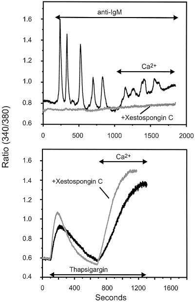
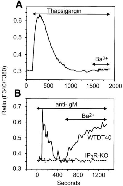
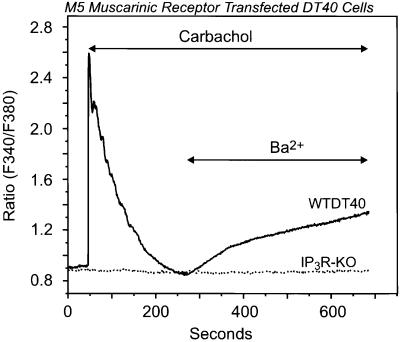
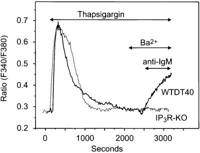
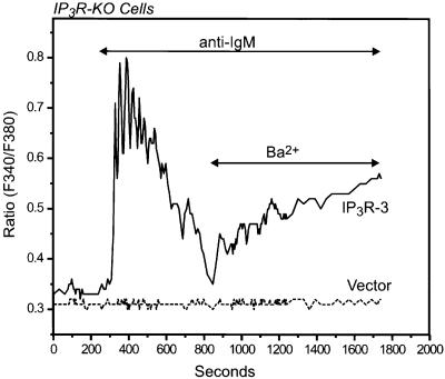
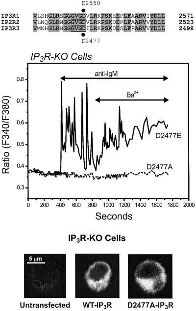
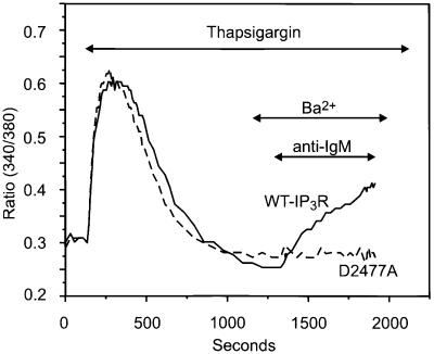
We examined the roles of inositol 1,4,5-trisphosphate (IP3) receptors (IP3R) in calcium signaling using DT40 B lymphocytes, and a variant lacking the three IP3R isoforms (IP3R-KO). In wild-type cells, B cell receptor (BCR) stimulation activates a cation entry route that exhibits significantly greater permeability to Ba2+ than does capacitative calcium entry. This cation entry is absent in IP3R-KO cells. Expression of the type-3 IP3R (IP3R-3) in the IP3R-KO cells rescued not only agonist-dependent release of intracellular Ca2+, but also Ba2+ influx following receptor stimulation. Similar results were obtained with an IP3R-3 mutant carrying a conservative point mutation in the selectivity filter region of the channel (D2477E); however, an IP3R-3 mutant in which this same aspartate was replaced by alanine (D2477A) failed to restore either BCR-induced Ca2+ release or receptor-dependent Ba2+ entry. These results suggest that in DT40 B lymphocytes, BCR stimulation activates a novel cation entry across the plasma membrane that depends upon, or is mediated by, fully functional IP3R.


Figures

None
Fig. 1. The IP3R inhibitor xestospongin C effectively suppresses both BCR-induced Ca2+ release and calcium entry in DT40 cells B lymphocytes, but does not affect thapsigargin-induced Ca2+ release or entry. DT40 cells were incubated for 45 min in the presence of 25 µM xestospongin C. Cells were then bathed in nominally Ca2+-free medium, exposed to 5 µg/ml anti-IgM antibody (top) or 2 µM thapsigargin (bottom), as indicated. Ca2+ (1.5 mM) was added where indicated. In this experiment, relatively higher ratio values were obtained as compared with other experiments due to the use of a different Ca2+ measuring system; in this series (and for experiments in Figure 3), a photon counting system was used, while in the other series, an imaging system was used. Black trace: control (no xestospongin C); gray trace, 25 µM xestospongin C. Shown are representative traces from three independent experiments.
None
Fig. 2. BCR activation, but not store depletion, induces Ba2+ entry in DT40 B lymphocytes. (A) Fura-2-loaded WTDT40 cells were incubated in nominally Ca2+-free medium and then exposed to 2 µM thapsigargin in order to deplete intracellular Ca2+ stores. After cytosolic Ca2+ returned to basal levels, Ba2+ (10 mM) was added to the medium. A representative trace from at least five independent experiments is shown. (B) Wild-type (WTDT40) or IP3R-KO (broken line) DT40 cells were maintained in nominally Ca2+-free medium, exposed to 5 µg/ml anti-IgM antibody, and then Ba2+ (10 mM) was added where indicated. Representative traces from at least five independent experiments are shown.
None
Fig. 3. Activation of the G-protein-coupled M5 muscarinic receptor results in stimulation of Ba2+ entry in wild-type but not in IP3R-KO DT40 cells. Ba2+ influx was measured in single Fura-2-loaded wild-type (WTDT40) or IP3R-KO (dotted line) DT40 cells transfected with the human M5 muscarinic receptor. The cells were maintained in nominally Ca2+-free medium, exposed to 100 µM carbachol, and then Ba2+ (10 mM) was added where indicated. The relatively higher ratio values in this experiment are due to the use of a different Ca2+ measuring system; in this series, a photon counting system was used, while in the other series, an imaging system was used. Representative traces from three independent experiments are shown.
None
Fig. 4. BCR-dependent activation of Ba2+ entry in wild-type but not in IP3R-KO DT40 cells. Fura-2-loaded wild-type (WTDT40) and IP3R-KO DT40 cells were incubated in nominally Ca2+-free medium and then exposed to 2 µM thapsigargin. After complete store depletion, Ba2+ (10 mM) was added to the medium where indicated, and 2 min later anti-IgM (5 µg/ml) was added. Representative traces from three independent experiments are shown.
None
Fig. 5. Transient expression of an IP3R restores agonist-induced Ba2+ entry in IP3R-KO DT40 cells. Ba2+ influx was measured in Fura-2-loaded IP3R-KO DT40 cells transfected with either the rat IP3R-3 or its vector (Mock). The cells were maintained in nominally Ca2+-free medium, exposed to 5 µg/ml anti-IgM antibody, and then Ba2+ (10 mM) was added where indicated. Representative traces from three independent experiments are shown.
None
Fig. 6. Effect of single point mutations within the putative pore forming region of IP3R-3 on restoration of agonist-induced Ba2+ entry in IP3R-KO DT40 cells. The top of the figure shows an alignment of a region including the predicted pore of rat IP3R-1–3 (the putative selectivity filter sequence is underlined). D2477 of rat IP3R-3, which was mutated into glutamate (D2477E) or alanine (D2477A), corresponds to D2550 of rat IP3R-1. In the experiment shown below, Ba2+ influx was measured in Fura-2-loaded IP3R-KO DT40 cells transfected with either the D2477E (solid line) or the D2477A (broken line) mutants of the rat IP3R-3 (see Materials and methods for details on the mutations). The cells were maintained in nominally Ca2+-free medium, exposed to 5 µg/ml anti-IgM antibody, and then Ba2+ (10 mM) was added where indicated. Representative traces from three independent experiments are shown. At the bottom, immunolocalization of transiently expressed rat IP3R-3 (pCB6+ vector) in DT40 B cells is shown. IP3R-KO cells untransfected, or transfected with the wild-type rat IP3R-3 (WT-IP3R), or the rat IP3R-3 mutant D2477A (D2477A-IP3R; see text for details) were incubated with a mouse anti-IP3R-3 monoclonal antibody and then with the secondary Alexa Fluor 488-labeled anti-mouse IgG antibody. The fluorescence images were acquired with a Zeiss 410 confocal microscope.
None
Fig. 7. The D2477A mutant of the rat IP3R-3 does not restore BCR-dependent activation of Ba2+ entry in Ca2+-depleted, IP3R-KO DT40 cells. Fura-2-loaded IP3R-KO DT40 cells transfected with either the wild type or the D2477A mutant of the rat IP3R-3 were incubated in nominally Ca2+-free medium and then exposed to 2 µM thapsigargin. After complete store depletion, Ba2+ (10 mM) was added to the medium where indicated, and 2 min later anti-IgM (5 µg/ml) was added. Solid line, wild-type IP3R; dashed line, D2477A IP3R. Representative traces from three independent experiments are shown.

References

    1. Baba T.W., Giroir,B.P. and Humphries,E.H. (1985) Cell lines derived from avian lymphomas exhibit two distinct phenotypes. Virology, 144, 139–151. - PubMed
    1. Barritt G.J. (1999) Receptor-activated Ca2+ inflow in animal cells: a variety of pathways tailored to meet different intracellular Ca2+ signalling requirements. Biochem. J., 337, 153–169. - PMC - PubMed
    1. Berridge M.J. (1993) Inositol trisphosphate and calcium signalling. Nature, 361, 315–325. - PubMed
    1. Berridge M.J. (1995) Capacitative calcium entry. Biochem. J., 312, 1–11. - PMC - PubMed
    1. Bezprozvanny I. and Ehrlich,B.E. (1994) Inositol (1,4,5)-trisphosphate (InsP3)-gated Ca channels from cerebellum: Conduction properties for divalent cations and regulation by intraluminal calcium. J. Gen. Physiol., 104, 821–856. - PMC - PubMed

MeSH terms