. 2002 Sep 2;21(17):4560-70.
doi: 10.1093/emboj/cdf438.

A novel cGMP signalling pathway mediating myosin phosphorylation and chemotaxis in Dictyostelium

Affiliations

A novel cGMP signalling pathway mediating myosin phosphorylation and chemotaxis in Dictyostelium

Leonard Bosgraaf et al. EMBO J. .

Abstract

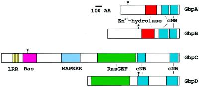
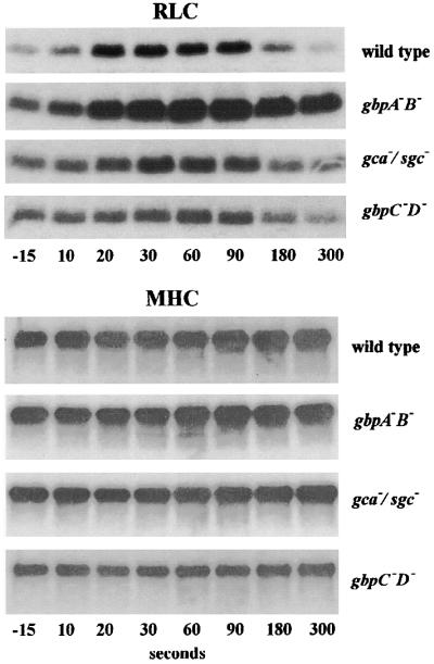
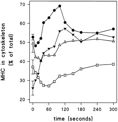
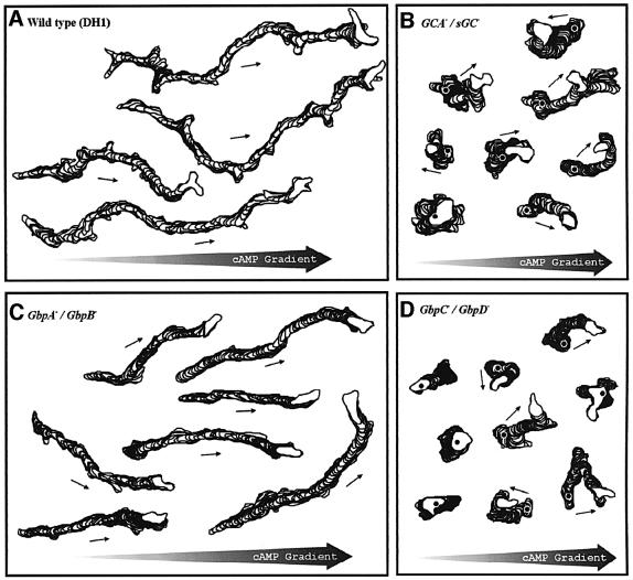
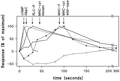
Chemotactic stimulation of Dictyostelium cells results in a transient increase in cGMP levels, and transient phosphorylation of myosin II heavy and regulatory light chains. In Dictyostelium, two guanylyl cyclases and four candidate cGMP-binding proteins (GbpA- GbpD) are implicated in cGMP signalling. GbpA and GbpB are homologous proteins with a Zn2+-hydrolase domain. A double gbpA/gbpB gene disruption leads to a reduction of cGMP-phosphodiesterase activity and a 10-fold increase of basal and stimulated cGMP levels. Chemotaxis in gbpA(-)B(-) cells is associated with increased myosin II phosphorylation compared with wild-type cells; formation of lateral pseudopodia is suppressed resulting in enhanced chemotaxis. GbpC is homologous to GbpD, and contains Ras, MAPKKK and Ras-GEF domains. Inactivation of the gbp genes indicates that only GbpC harbours high affinity cGMP-binding activity. Myosin phosphorylation, assembly of myosin in the cytoskeleton as well as chemotaxis are severely impaired in mutants lacking GbpC and GbpD, or mutants lacking both guanylyl cyclases. Thus, a novel cGMP signalling cascade is critical for chemotaxis in Dictyostelium, and plays a major role in myosin II regulation during this process.


Figures

None
Fig. 1. Four unusual putative cGMP-binding proteins in Dictyostelium. Schematic of the topology of GbpA–GbpD. The positions of disruption of the protein sequences in the knock-out cell lines are indicated by asterisks. The bar refers to 100 amino acids. GbpA and GbpB have the same domain topology; a Zn2+-hydrolase (red) and two cNB domains (blue). GbpC and GbpD are also homologues, sharing an N-terminal Ras-GEF-associated domain (light green) a Ras-GEF domain (green), a GRAM domain (dark blue) and two cNB domains (blue). GbpC has additional N-terminal sequence containing leucine-rich repeats (yellow), a Ras (orange), a MAP kinase kinase kinase (pink) and a DEP (turquoise) domain.
None
Fig. 2. cGMP response in gbp-null cells. The cell were starved for 5 h followed by stimulation with 0.1 µM cAMP. Responses were terminated with perchloric acid, and cGMP levels were measured. (A) The symbols refer to wild-type DH1 (filled circles); gbpA (open triangles); gbpB (open squares); and gbpAB (closed squares). (B) Wild-type DH1 (filled circles); gbpC (open inverted triangles); gbpD (open circles); and gbpCD (filled inverted triangles). Identical data are presented for wild-type DH1 in (A) and (B). The results shown are the means of triplicate determinations from a typical experiment repeated once.
None
Fig. 3. cAMP and cGMP binding to proteins from gbp-null cells. The binding of 10 nM [3H]cAMP (A) or 10 nM [3H]cGMP (B) to the cytosolic fraction of wild-type DH1 and different null cell lines was measured. The means and standard deviations of two (A) or three (B) experiments with triplicate determinations are shown.
None
Fig. 4. Scatchard plot of cGMP binding to proteins from gbp-null cells. The binding of different concentrations of [3H]cGMP to the cytosolic fraction was determined. The symbols refer to wild-type DH1 (filled circles); gbpAB (filled squares); gbpC (open inverted triangles); gbpD (open circles); and gbpCD (filled inverted triangles). Curve fitting with a two-component model indicates two binding forms in wild-type, gbpAB and gbpD showing 200 ± 35 fmol high affinity cGMP-binding sites/mg protein with a Kd of 3.9 ± 1.5 nM and 1165 ± 134 fmol low affinity cGMP-binding sites/mg protein with a Kd of 257 ± 83 nM. Mutant gbpC and double mutant gbpCD show only a low affinity cGMP binding component with 1235 ± 333 fmol cGMP-binding sites/mg protein with a Kd of 557 ± 220 nM. The data points shown are the means of two experiments with triplicate determinations; the estimated binding constants are the means and standard error of the data pooled from three cell lines (wild-type, gbpAB and gbpD) or two cell lines (gbpC and gbpCD).
None
Fig. 5. cAMP-induced phosphorylation of myosin heavy chain II (MHC) and regulatory light chain (RLC). Starved cells were incubated with [32P]phosphate for 30 min, followed by stimulation with 1 μM cAMP at t = 0 s. Samples were taken at the times indicated and immunoprecipitated with antibodies against RLC and MHC. The immunoprecipitates were analysed by SDS–PAGE and autoradiography. Typical experiments are presented and were repeated at least once. A quantitative analysis of these data is presented in Table II.
None
Fig. 6. Association of MHC with the Triton-insoluble cytoskeleton. Starved cells were stimulated with 1 μM cAMP at t = 0 s. At the times indicated, 0.5% Triton was added, and lysates were separated into a supernatant and a Triton-insoluble cytoskeleton. The levels of MHC were determined by western blotting using antibody against MHC. The means of two or three experiments are shown. A quantitative analysis of these data is presented in Table II. The symbols refer to wild-type DH1 (filled circles); guanylyl cyclase gca/sgc null cells (open triangles); cGMP phosphodiesterase gbpAB null cells (filled inverted triangles); and gbpCD null cells lacking two cGMP targets (open squares).
None
Fig. 7. Cell movement in cAMP gradients. Aggregation-competent cells were deposited in a chemotaxis chamber with a cAMP gradient pointing to the right. Cells were videorecorded for 10 min at a rate of 15 frames per min. The cell perimeters were outlined and are presented for five representative cells as a stack of successive outlines at 8 s interval. The first outline is indicated by an asterisk. (A) Wild-type DH1 cells; (B) guanylyl cyclase gca/sgc null cells; (C) cGMP phosphodiesterase gbpAB null cells; and (D) gbpCD cells lacking the two cGMP targets.
None
Fig. 8. Time course of the cGMP and myosin responses in wild-type cells. The responses were calculated by first subtracting basal levels of unstimulated cells and are then expressed as a percentage of the maximal response. Data are from Figures 2, 5 and 6; the cGMP occupancy of GbpC is taken from Van Haastert et al. (1982a). Symbols refer to cGMP (open circles); phosphorylation of RLC (filled triangles); phosphorylation of MHC (filled inverted triangles); MHC in the cytoskeleton of wild-type cells (filled squares); and MHC in the cytoskeleton of gca/sgc and gbpCD cells (open squares). The cGMP occupancy of GbpC is presented as a dotted line.

References

    1. Berlot C.H., Devreotes,P.N. and Spudich,J.A. (1987) Chemoattractant-elicited increases in Dictyostelium myosin phosphorylation are due to changes in myosin localization and increases in kinase activity. J. Biol. Chem., 262, 3918–3926. - PubMed
    1. Boy-Marcotte E. and Jacquet,M. (1982) A Dictyostelium discoideum DNA fragment complements a Saccharomyces cerevisiae ura3 mutant. Gene, 20, 433–439. - PubMed
    1. Carfi A., Pares,S., Duee,E., Galleni,M., Duez,C., Frere,J.M. and Dideberg,O. (1995) The 3-D structure of a zinc metallo-β-lactamase from Bacillus cereus reveals a new type of protein fold. EMBO J., 14, 4914–4921. - PMC - PubMed
    1. Chandrasekhar A., Wessels,D. and Soll,D.R. (1995) A mutation that depresses cGMP phosphodiesterase activity in Dictyostelium affects cell motility through an altered chemotactic signal. Dev. Biol., 169, 109–122. - PubMed
    1. de la Roche M.A. and Cote,G.P. (2001) Regulation of Dictyostelium myosin I and II. Biochim. Biophys. Acta, 1525, 245–261. - PubMed

Publication types

MeSH terms