. 2002 Sep 2;21(17):4571-82.
doi: 10.1093/emboj/cdf446.

Tyrosine 221 in Crk regulates adhesion-dependent membrane localization of Crk and Rac and activation of Rac signaling

Affiliations

Tyrosine 221 in Crk regulates adhesion-dependent membrane localization of Crk and Rac and activation of Rac signaling

Yama A Abassi et al. EMBO J. .

Abstract

The adaptor protein CrkII plays a central role in signal transduction cascades downstream of a number of different stimuli. We and others have previously shown that CrkII mediates attachment-induced JNK activation, membrane ruffling and cell motility in a Rac-dependent manner. We report here that cell attachment leads to tyrosine phosphorylation of CrkII on Y221, and that CrkII-Y221F mutant demonstrates enhanced association with the Crk-binding partners C3G and paxillin. Despite this enhanced signaling complex formation, CrkII-Y221F fails to induce JNK and PAK activation, membrane ruffling and cell migration, suggesting that it is defective in activating Rac signaling. Wild-type CrkII has no effect on adhesion-induced GTP loading of Rac, but its expression results in enhanced membrane localization of Rac, which is known to be required for Rac signaling. In contrast, CrkII-Y221F is deficient in enhancing membrane localization of Rac. Mutations in Rac and CrkII-Y221F that force membrane targeting of these molecules restore Rac signaling in adherent cells. Together, these results indicate that the Y221 site in CrkII regulates Rac membrane translocation upon cell adhesion, which is necessary for activation of downstream Rac signaling pathways.


Figures

None
Fig. 1. Adhesion-dependent tyrosine phosphorylation of CrkII on Y221. (A) Serum-starved COS-7 cells were either held in suspension for 1 h and lysed (S) or held in suspension for 1 h and plated on FN-coated dishes for the indicated periods of time (in minutes), and cell lysates were immunoprecipitated with anti-Crk antibody, followed by immunoblotting with anti-phosphotyrosine antibody (‘anti-pY’, upper panel). The membranes were stripped and reprobed with anti-Crk antibodies (lower panel). (B) Lysates prepared from cells that had been either held in suspension as above or plated on FN for 30 min were subjected to immunoprecipitation with anti-pY221 antibody, which recognizes CrkII that is phosphorylated on Y221, followed by immunoblotting with anti-Crk antibodies (upper panel). Total cell lysate samples were immunoblotted with anti-Crk antibodies to ensure that equal amounts of CrkII protein were used for immunoprecipitation (lower panel).
None
Fig. 2. CrkII-Y221F mutant demonstrates enhanced interaction with paxillin. (A) COS-7 cells transfected with 3 µg of either wild-type CrkII or CrkII-Y221F mutant were serum starved and either held in suspension (S) for 1 h or held in suspension for 1 h and plated on FN-coated dishes for the indicated periods of time. The cells were lysed and immunoprecipitated with anti-Crk antibody, followed by immunoblotting with anti-phosphotyrosine, anti-paxillin and anti-Crk antibodies. (B) Quantitative analysis of the interactions between CrkII and paxillin was carried out as described in Materials and methods. Interaction observed in wild-type CrkII-expressing cells in suspension was arbitrarily defined as ‘1’. *P < 0.01 between wild-type CrkII and CrkII-Y221F samples in suspension and at 15 and 30 min time points; **P < 0.05 between wild-type CrkII and CrkII-Y221F samples at 60 min time point. Results are shown for experiments independently carried out three times.
None
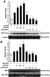
Fig. 3. CrkII-Y221F mutant negatively regulates Crk signaling upon cell attachment. COS-7 cells were transiently transfected with the indicated amounts (in µg) of wild-type CrkII or CrkII-Y221F mutant, together with 0.25 µg of HA-tagged JNK (A) or myc-tagged PAK (B). The cells were then held in suspension (white bar) for 5 min [in (A)] or for 3 h [in (B)] and then plated on FN (black bars) for 15 min. Kinase assays, densitometric and statistical analysis were carried out as described in Materials and methods. *P < 0.01 between the indicated samples and HA-JNK/Myc-PAK alone-transfected cells plated on FN; **P < 0.005 between the indicated samples and HA-JNK/Myc-PAK alone-transfected cells plated on FN. (C) COS-7 cells were transiently transfected with 0.5 µg of GFP (‘Control’), wild-type GFP–CrkII or GFP–CrkII-Y221F constructs. The cells were subjected to a haptotactic migration assay on FN as described in Materials and methods. The cells that had migrated to the underside of the Transwell membrane were fixed, and GFP-positive cells were visualized and photographed. At least six different fields were counted per membrane and the results were normalized to the transfection efficiency. In (A) and (B), results are shown for experiments independently carried out three times. In (C), bars indicate SD in a representative experiment carried out in triplicate.
None
Fig. 4. Expression of CrkII does not affect GTP loading of Rac in FN-adherent COS-7 cells. COS-7 cells were transiently transfected with the indicated amounts (in µg) of wild-type CrkII, Crk-Y221F mutant or wild-type Vav2, together with 0.25 µg of myc-tagged wild-type Rac. The cells were held in suspension for 3 h, plated on FN for 15 min (black bars) or held in suspension for a further 15 min (white bar) and lysed in PBD lysis buffer. Cell lysates were incubated with GST–PBD beads, and bound GTP-Rac as well as the amount of myc-Rac in the lysate was detected by anti-myc immunoblotting. The amount of GTP-Rac signal in each sample was normalized to the amount of total myc-Rac expressed in the sample. Results are shown for experiments independently carried out three times.
None
Fig. 5. Subcellular localization of Rac and CrkII in wild-type CrkII- and CrkII-Y221F mutant-expressing cells. (A) Wild-type CrkII or CrkII-Y221F mutant was co-expressed with GFP-tagged wild-type Rac in COS-7 cells. The cells were detached and reattached on FN-coated coverslips for 15 min, followed by staining with anti-Crk antibodies. The cells were visualized and photographed by confocal microscopy. (B) COS-7 cells expressing myc-Rac or myc-Rac with wild-type CrkII or CrkII-Y221F were serum starved, detached and reattached to FN-coated dishes. The cells were fractionated as described in Materials and methods in order to obtain cytosolic and membrane fractions. Equal amounts of protein from these fractions were loaded on a gel and immunoblotted with anti-Rac or anti-Crk antibodies. (C) The bands corresponding to myc-Rac were densitometrically scanned and the ratio of the relative intensity is shown. The data are representative of three independent experiments. *P < 0.005 between wild-type CrkII and control samples.
None
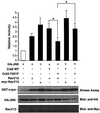
Fig. 6. CrkII-Y221F inhibits RacV12-, but not myr-RacV12-induced JNK activation upon cell attachment. COS-7 were transiently transfected with 0.25 µg of plasmids encoding either wild-type CrkII or CrkII-Y221F mutant, or 0.1 µg of plasmids coding for RacV12 or myr-RacV12 together with 0.25 µg of HA-JNK, as indicated. Transfected cells were detached and reattached on FN-coated dishes for 15 min. Cell lysates were subjected to immunoprecipitation with anti-HA antibodies, followed by immunocomplex kinase assay, as described in Materials and methods. *P < 0.05 between CrkII-Y221F/RacV12 and wild-type CrkII/RacV12 samples, and between CrkII-Y221F/myr-RacV12 and CrkII-Y221F/RacV12 samples. Results are shown for experiments independently carried out three times.
None
Fig. 7. Subcellular localization of RacV12 and myr-RacV12 in cells expressing wild-type CrkII and CrkII-Y221F mutant. Wild-type CrkII or CrkII-Y221F was co-expressed with GFP-tagged RacV12 in (A), and GST-tagged CrkII or GST-tagged CrkII-Y221F was expressed with myc-tagged myr-RacV12 in (B) in COS-7 cells. The cells were detached, replated on FN-coated coverslips for 15 min, and stained with the indicated antibodies. The cells were visualized and photographed by confocal microscopy.
None
Fig. 8. Membrane-targeted myr-CrkII-Y221F induces membrane localization of wild-type Rac and RacV12. COS-7 cells transiently expressing myr-CrkII-Y221F along with GFP–Rac and GFP–RacV12 were plated on FN-coated chamber slides for 15 min, followed by staining with anti-Crk antibodies. The cells were visualized and photographed using confocal microscopy.
None
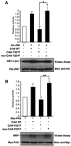
Fig. 9. Membrane-targeted myr-CrkII-Y221F induces activation of JNK and PAK. COS-7 cells were transiently transfected with 0.25 µg of the indicated CrkII constructs together with 0.25 µg of HA-tagged JNK (A) or myc-tagged PAK (B). The cells were held in suspension for 5 min (A) or 3 h (B) and then plated on FN for 15 min. Kinase assays and densitometric analysis were carried out as described in Materials and methods. *P < 0.01 between CrkII-Y221F and myr-CrkII-Y221F samples; **P < 0.025 between Crk-Y221F and myr-CrkII-Y221F samples. (C) Serum-starved COS-7 cells expressing myc-Rac with or without wild-type CrkII or myr-CrkII-Y221F were detached, held in suspension for 3 h and plated on FN-coated dishes for 15 min. The cells were lysed and analyzed for Rac-GTP loading as described in Materials and methods. Results are shown for independent experiments carried out three times.

References

    1. Abe K., Rossman,K.L., Liu,B., Ritola,K.D., Chiang,D., Campbell,S.L., Burridge,K. and Der,C.J. (2000) Vav2 is an activator of Cdc42, Rac1 and RhoA. J. Biol. Chem., 275, 10141–10149. - PubMed
    1. Anafi M., Rosen,M.K., Gish,G.D., Kay,L.E. and Pawson,T. (1996) A potential SH3 domain-binding site in the Crk SH2 domain. J. Biol. Chem., 271, 21365–21374. - PubMed
    1. Bagrodia S., Derijard,B., Davis,R.J. and Cerione,R.A. (1995) Cdc42 and PAK-mediated signaling leads to Jun kinase and p38 mitogen-activated protein kinase activation. J. Biol. Chem., 270, 27995–27998. - PubMed
    1. Beitner-Johnson D. and LeRoith,D. (1995) Insulin-like growth factor-I stimulates tyrosine phosphorylation of endogenous c-Crk. J. Biol. Chem., 270, 5187–5190. - PubMed
    1. Benard V., Bohl,B.P. and Bokoch,G.M. (1999) Characterization of rac and cdc42 activation in chemoattractant-stimulated human neutrophils using a novel assay for active GTPases. J. Biol. Chem., 274, 13198–13204. - PubMed

Publication types

MeSH terms