. 2002 Sep 2;21(17):4632-40.
doi: 10.1093/emboj/cdf460.

The chromatin remodeling complex NoRC targets HDAC1 to the ribosomal gene promoter and represses RNA polymerase I transcription

Affiliations

The chromatin remodeling complex NoRC targets HDAC1 to the ribosomal gene promoter and represses RNA polymerase I transcription

Yonggang Zhou et al. EMBO J. .

Abstract

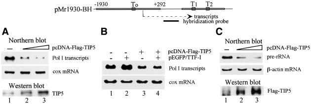
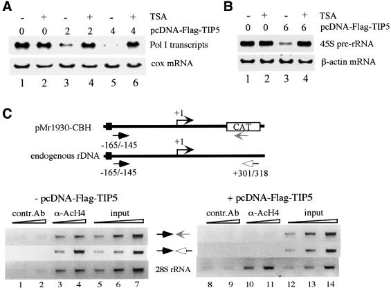
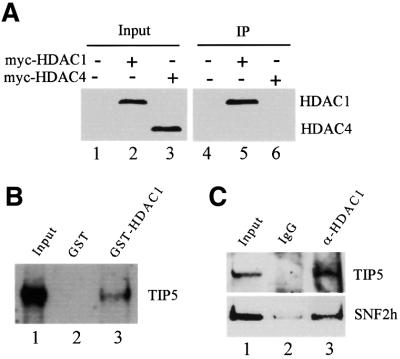
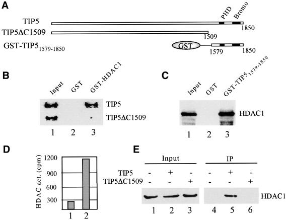
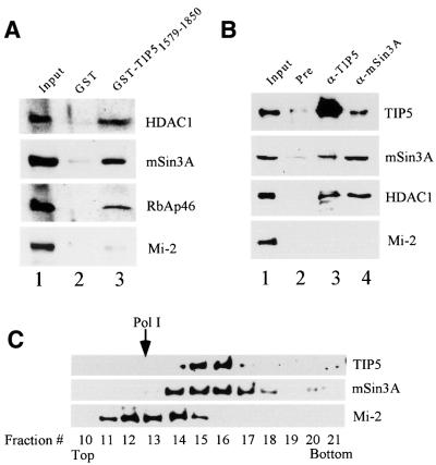
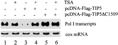
Mammalian chromatin remodeling complexes are involved in both activation and repression of transcription. Here, we show that NoRC, a SNF2h- containing nucleolar chromatin remodeling complex, represses ribosomal gene transcription. NoRC-mediated rDNA silencing was alleviated by trichostatin A, indicating that histone deacetylation is causally involved in silencing. Chromatin immunoprecipitation experiments demonstrate that overexpression of TIP5, the large subunit of NoRC, mediates deacetylation of nucleosomes in the vicinity of the rDNA promoter. Protein-protein interaction assays reveal association of TIP5 with the histone deacetylase HDAC1 in vivo and in vitro. Deletion of the C-terminal PHD finger and bromodomain abolishes the interaction of TIP5 and HDAC1, and abrogates transcriptional repression. The results suggest that NoRC silences the rDNA locus by targeting the SIN3 corepressor complex to the rDNA promoter, thereby establishing a repressed chromatin structure.


Figures

None
Fig. 1. Overexpression of TIP5 inhibits Pol I transcription in vivo. (A) TIP5 represses Pol I transcription in vivo. NIH 3T3 cells were transiently transfected with 2.5 µg of Pol I reporter (pMr1930-BH) and 2 µg (lane 2) or 4 µg (lane 3) of pcDNA-Flag-TIP5. Total RNA was extracted and Pol I transcripts were monitored on northern blots. As an internal control, the amount of cytochrome c oxidase (cox) mRNA was determined. Overexpression of TIP5 was visualized on western blots using α-TIP5 antibodies. A schematic representation of the Pol I reporter and the hybridization probe used in northern blots is shown above. (B) TIP5 counteracts transcription activation of TTF-I in vivo. NIH 3T3 cells were transfected with 2.5 µg of Pol I reporter, 2 µg of pEGFP/TTF-I and 4 µg of pcDNA-Flag-TIP5 as indicated. Total RNA was extracted and transcripts from the Pol I reporter were monitored on northern blots. To normalize for variations of RNA loading, the blot was also hybridized with a probe complementary to cox mRNA. (C) Overexpression of TIP5 inhibits cellular 45S pre-rRNA synthesis. 293T cells were transiently transfected with pcDNA-Flag-TIP5, and RNA was analyzed on northern blots using a riboprobe complementary to nucleotides 1–155 of human pre-rRNA. The blots were subsequently reprobed for actin mRNA. Overexpression of TIP5 was visualized on western blots using α-Flag antibodies.
None
Fig. 2. Transcription repression by TIP5 is mediated by histone deacetylation. (A) TSA counteracts TIP5-mediated repression of Pol I transcription. NIH 3T3 cells were co-transfected with 2.5 µg of Pol I reporter (pMr1930-BH) and 2 µg (lanes 3 and 4) or 4 µg (lanes 5 and 6) of pcDNA-Flag-TIP5. Where indicated, TSA (33 nM) was added 24 h after transfection, and cells were cultured for a further 24 h. Transcripts from the Pol I reporter and endogenous cox gene were monitored on northern blots. (B) TIP5-mediated repression of endogenous rDNA transcription is alleviated by TSA. 293T cells were transfected with 6 µg of pcDNA-Flag-TIP5 and cultured in the absence or presence of 33 nM TSA as indicated. 45S pre-rRNA synthesis was monitored on northern blots. (C) ChIP assay. NIH 3T3 cells were co-transfected with 2.5 µg of Pol I reporter (pMr1930-CBH) in the absence (lanes 1–7) or presence (lanes 8–14) of 4 µg of pcDNA-Flag-TIP5. After formaldehyde cross-linking, cells were sonicated and soluble chromatin was immunoprecipitated with anti-AcH4 (lanes 3, 4, 10 and 11) or anti-myc antibodies (lanes 1, 2, 8 and 9). Different amounts of precipitated DNA were amplified by PCR. Primers were used that amplify either the promoter of the reporter plasmid or the endogenous rDNA (as indicated in the scheme above) or part of the 28S rRNA coding region (lower). Quantitative PCR analysis was also performed before immunoprecipitation to ensure equal transfection efficiencies (input).
None
Fig. 3. TIP5 interacts with HDAC1 in vitro and in vivo. (A) HDAC1 but not HDAC4 co-immunoprecipitates with TIP5. pcDNA-myc-HDAC1 or pcDNA-myc-HDAC4 was co-transfected with pcDNA-Flag-TIP5 into 293T cells. Cells were lysed and NoRC was precipitated with anti-Flag antibodies. Co-immunoprecipitated HDAC1 and HDAC4 were monitored on western blots with α-myc antibodies. The amount of HDAC1 and HDAC4 present in 5% of the cell lysates is shown in lanes 1–3. (B) TIP5 interacts with HDAC1. A total of 100 µl of a partially purified nuclear extract (DEAE-280 fraction) were applied to 10 µl of glutathione–Sepharose beads containing equivalent amounts of immobilized GST (lane 2) or GST–HDAC1 (lane 3). After washing with buffer AM-300, bound proteins (50%) were analyzed on western blots using anti-TIP5 antibodies. Ten percent of input of the DEAE-280 fraction is shown in lane 1. (C) NoRC interacts with HDAC1 in vivo. Pre-immune serum (lane 2) or anti-HDAC1 antibodies (lane 3) were bound to protein G–agarose, and 10 µl of beads were incubated with 100 µl of DEAE-280 fraction at 4°C for 4 h. Captured NoRC complex was analyzed on western blots using anti-TIP5 and SNF2h antibodies. Ten percent of the DEAE-280 fraction (lane 1) and 50% of bead-bound proteins are shown in lanes 2 and 3.
None
Fig. 4. The C-terminal part of TIP5 interacts with HDAC1. (A) Schematic representation of TIP5, TIP5ΔC1509 and GST–TIP51579–1850. The position of the PHD finger and bromodomain is indicated. (B) HDAC1 interacts with the C-terminal region of TIP5. A total of 10 µl of immobilized GST (lane 2) or GST–HDAC1 (lane 3) were incubated with 5 µl of 35S-labeled TIP5 and TIP5ΔC1509 in 200 µl of buffer AM-100. Bound proteins (50%) were analyzed by electrophoresis and autoradiography. Ten percent of input proteins are shown in lane 1. (C) The C-terminal part of TIP5 including the PHD finger and bromodomain interacts with HDAC1. A total of 10 µl of bead-bound GST (lane 2) or GST–TIP51579–1850 (lane 3) were incubated with 5 µl of 35S-labeled HDAC1, and captured HDAC1 was analyzed by 10% SDS–PAGE and autoradiography. Ten percent of input is shown in lane 1. (D) The C-terminal part of TIP5 recruits HDAC activity. Bead-bound GST and GST–TIP51579–1850 were incubated with 200 µl of nuclear extracts for 4 h at 4°C. Washed beads were assayed for the release of [3H]acetate (c.p.m.) from 3H-acetylated histones. (E) Interaction of TIP5 and HDAC1 in vivo. 293T cells were co-transfected with 4 µg of pcDNA-myc-HDAC1 and 8 µg of pcDNA-Flag-TIP5 or pcDNA-Flag-TIP5ΔC1509. TIP5-containing protein complexes were precipitated with α-Flag antibodies, and the presence of HDAC1 in the immunoprecipitates was analyzed on western blots with α-myc antibodies. Five percent of input is shown in lanes 1–3.
None
Fig. 5. NoRC interacts with the SIN3 deacetylase complex. (A) TIP5 interacts with the SIN3 complex in vitro. A total of 100 µl of a DEAE-280 fraction were incubated with 10 µl of immobilized GST (lane 2) or GST–TIP51579–1850 (lane 3). After washing with buffer AM-300, bound proteins were analyzed on western blots using α-HDAC1, α-mSin3A, α-RbAp46 and α-Mi-2 antibodies. Ten percent of input is shown in lane 1. (BIn vivo association of NoRC with the SIN3 complex. A total of 100 µl of a DEAE-280 fraction were incubated with pre-immune serum (lane 2), α-TIP5 (lane 3) and α-mSin3A antibodies (lane 4) bound to protein G–agarose. After washing with buffer AM-200, 50% of bead-bound proteins were analyzed on western blots using antibodies against HDAC1, TIP5, mSin3A and Mi-2. Ten percent of input proteins are shown in lane 1. (C) Co-sedimentation of NoRC with cellular SIN3. A total of 250 µl of a DEAE-280 fraction were centrifuged through a 12.5–30% glycerol gradient. Fractions (150 µl) were precipitated with trichloroacetic acid and analyzed on immunoblots using α-TIP5, α-Sin3A and α-Mi-2 antibodies. The position of Pol I (∼600 kDa) is marked by an arrow.
None
Fig. 6. Overexpression of a TIP5 C-terminal deletion mutant enhances rDNA transcription. NIH 3T3 cells were transfected with 2.5 µg of Pol I reporter (pMr1930-BH) and 4 µg of either pcDNA-Flag-TIP5 or pcDNA-Flag-TIP5ΔC1509. Where indicated, cells were treated with TSA (33 nM) for 24 h. Pol I transcripts and cox mRNA were visualized on northern blots.

References

    1. Ahringer J. (2000) NuRD and SIN3 histone deacetylase complexes in development. Trends Genet., 16, 351–356. - PubMed
    1. Ayer D. (1999) Histone deacetylases: transcriptional repression with SINers and NuRDs. Trends Cell Biol., 9, 193–198. - PubMed
    1. Bozhenok L., Wade,P.A. and Varga-Weisz,P. (2002) WSTF–ISWI chromatin remodeling complex targets heterochromatic replication foci. EMBO J., 21, 2231–2241. - PMC - PubMed
    1. Brehm A., Nielsen,S.J., Miska,E.A., McCance,D.J., Reid,J.L., Bannister,A.J. and Kouzarides,T. (1999) The E7 oncoprotein associates with Mi2 and histone deacetylase activity to promote cell growth. EMBO J., 18, 2449–2458. - PMC - PubMed
    1. Brown C.J. and Robinson,W.P. (2000) The causes and consequences of random and non-random X chromosome inactivation in humans. Clin. Genet., 58, 353–363. - PubMed

Publication types

MeSH terms