. 2002 Sep 2;21(17):4663-70.
doi: 10.1093/emboj/cdf476.

MicroRNA maturation: stepwise processing and subcellular localization

Affiliations

MicroRNA maturation: stepwise processing and subcellular localization

Yoontae Lee et al. EMBO J. .

Abstract

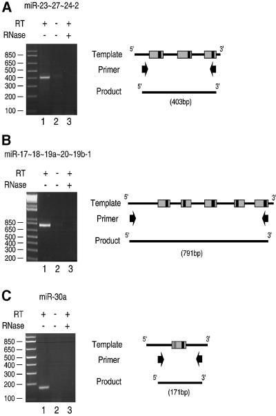
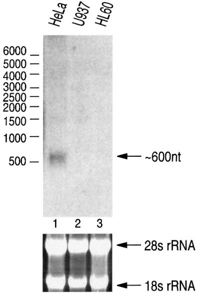
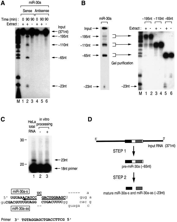
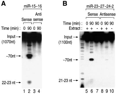
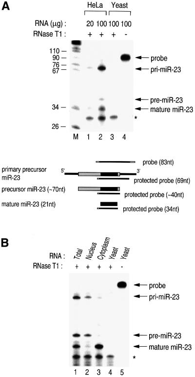
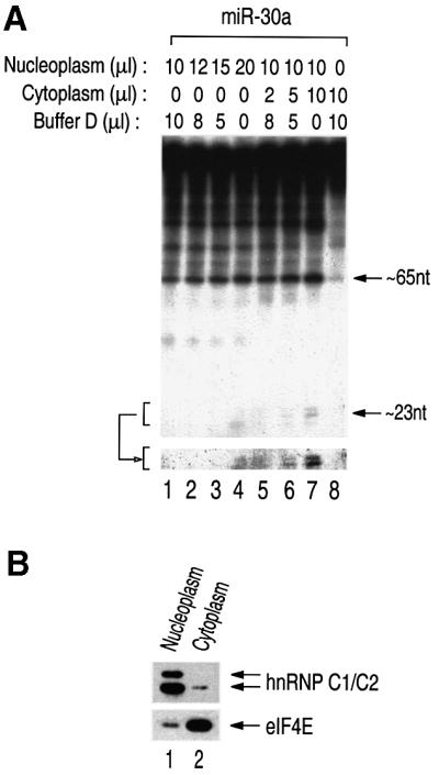
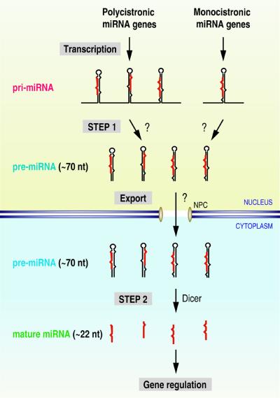
MicroRNAs (miRNAs) constitute a novel, phylogenetically extensive family of small RNAs ( approximately 22 nucleotides) with potential roles in gene regulation. Apart from the finding that miRNAs are produced by Dicer from the precursors of approximately 70 nucleotides (pre-miRNAs), little is known about miRNA biogenesis. Some miRNA genes have been found in close conjunction, suggesting that they are expressed as single transcriptional units. Here, we present in vivo and in vitro evidence that these clustered miRNAs are expressed polycistronically and are processed through at least two sequential steps: (i) generation of the approximately 70 nucleotide pre-miRNAs from the longer transcripts (termed pri-miRNAs); and (ii) processing of pre-miRNAs into mature miRNAs. Subcellular localization studies showed that the first and second steps are compartmentalized into the nucleus and cytoplasm, respectively, and that the pre-miRNA serves as the substrate for nuclear export. Our study suggests that the regulation of miRNA expression may occur at multiple levels, including the two processing steps and the nuclear export step. These data will provide a framework for further studies on miRNA biogenesis.


Figures

None
Fig. 1. RT–PCR reveals the long transcripts containing miRNAs. (A) HeLa total RNA was used for reverse transcription using a primer that binds downstream of mir-23∼27∼24-2. The product from the reverse transcription reaction served as the template for PCR to amplify the indicated regions containing the miRNA cluster. As a control, reverse transcriptase was omitted (lane 2). For RNase treatment, DNase-free RNase A (Ambion) was added to a final concentration of 50 µg/ml and incubated at 37°C for 2 min before reverse transcription. The illustration on the right side shows the template miRNA genes, the primers for PCR and the PCR product. Gray and black boxes indicate the regions corresponding to ∼70 nt precursors and mature miRNAs, respectively. (B) A similar experiment as in (A) was performed for mir-17∼18∼19a∼20∼19b-1. (C) A similar experiment as in (A) was performed for mir-30a.
None
Fig. 2. Northern blot analysis of miR-30a. From HeLa total RNA, a band of ∼600 nt was detected when 20 µg of RNA were analyzed on 1% formaldehyde–agarose gel and the blot was probed with miR-30a-specific probe. The size was determined by using RNA Millennium size markers from Ambion. The lower panel shows ethidium bromide staining of the agarose gel.
None
Fig. 3. In vitro processing of miRNAs via two sequential steps. (A) Uniformly radiolabeled RNAs corresponding to 371 nt covering miR-30a were incubated either in the presence or absence of HEK293T cell extract. Following the indicated incubation time, RNA was extracted and analyzed on a 12.5% (v/v) denaturing polyacrylamide gel. The marker in the left lane is pBR322 digested with MspI and end-labeled with T4 polynucleotide kinase. (B) Each fragment from the in vitro processing reaction was gel purified and incubated with cell extract for 90 min. (C) Primer extension analysis. From both the HeLa total RNAs and the in vitro processing product, ∼23 nt bands were detected using the 5′-end-labeled antisense oligonucleotide to miR-30a-s. (D) Schematic representation of in vitro processing.
None
Fig. 4. The in vitro processing system is a general analysis tool for miRNA maturation. (AIn vitro processing of miR-15∼16. These clustered miRNAs can also be processed in vitro to result in ∼70 nt intermediates and 22–23 nt final products. (BIn vitro processing of miR-23∼27∼24-2.
None
Fig. 5. Subcellular localization of the three different forms of miRNA. (A) RPA to detect miRNA. The indicated amounts of RNA from either HeLa or yeast cells were used for RPA. The asterisk indicates the self-protected probe. Schematically represented are the probes for the RPA and the protected fragments that are expected for each miRNA species. (B) Total, nuclear or cytoplasmic RNA from HeLa cells or yeast RNA was used for RPA.
None
Fig. 6. Nucleocytoplasmic compartmentalization of miRNA processing. (AIn vitro processing of miRNA in fractionated HeLa cell extract. The indicated amounts of the nuclear or cytoplasmic fraction were used for in vitro processing. The lower panel shows the ∼22 nt fragments after longer exposure. (B) Western blot analysis. Nucleoplasmic and cytoplasmic fractions were probed with anti-hnRNP C1/C2 antibody and anti-eIF4E antibody.
None
Fig. 7. A model for miRNA biogenesis. miRNA genes are transcribed by an unidentified polymerase to generate the primary transcripts, referred to as pri-miRNAs. Illustrated in the upper left is the clustered miRNA, such as miR-23∼27∼24-2, of which the pri-miRNA is polycistronic. Illustrated in the upper right is the miRNA, such as miR-30a, of which the pri-miRNA is monocistronic. The first-step processing (STEP 1) by an unknown factor(s) results in pre-miRNAs of ∼70 nt, which are recognized and exported by an unidentified export receptor. Upon export, Dicer, and possibly other factors, participates in the second-step processing (STEP 2) to produce mature miRNAs. The final product may function in a variety of regulatory pathways, such as translational control of certain mRNAs. The question marks indicate unidentified factors.

References

    1. Bernstein E., Caudy,A.A., Hammond,S.M. and Hannon,G.J. (2001) Role for a bidentate ribonuclease in the initiation step of RNA interference. Nature, 409, 363–366. - PubMed
    1. Chang D.D. and Sharp,P.A. (1989) Regulation by HIV Rev depends upon recognition of splice sites. Cell, 59, 789–795. - PubMed
    1. Conti E. and Izaurralde,E. (2001) Nucleocytoplasmic transport enters the atomic age. Curr. Opin. Cell Biol., 13, 310–319. - PubMed
    1. Dreyfuss G., Kim,V.N. and Kataoka,N. (2002) Messenger-RNA-binding proteins and the messages they carry. Nat. Rev. Mol. Cell Biol., 3, 195–205. - PubMed
    1. Fornerod M., Ohno,M., Yoshida,M. and Mattaj,I.W. (1997) CRM1 is an export receptor for leucine-rich nuclear export signals. Cell, 90, 1051–1060. - PubMed

Publication types