

Tissue localization of the endosymbiotic bacterium "Candidatus Blochmannia floridanus" in adults and larvae of the carpenter ant Camponotus floridanus
- PMID: 12200264
- PMCID: PMC124124
- DOI: 10.1128/AEM.68.9.4187-4193.2002
Tissue localization of the endosymbiotic bacterium "Candidatus Blochmannia floridanus" in adults and larvae of the carpenter ant Camponotus floridanus
Abstract
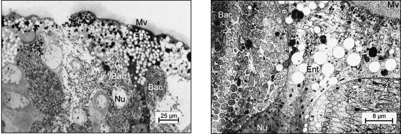
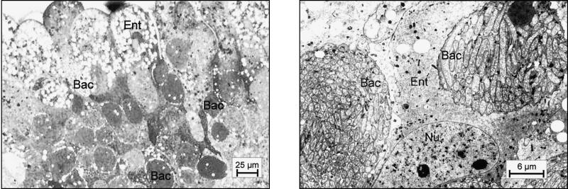
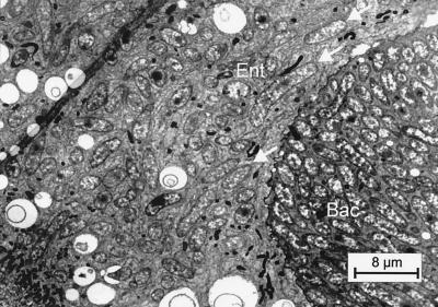
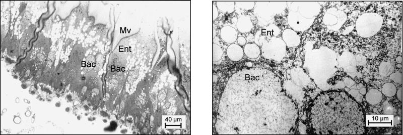
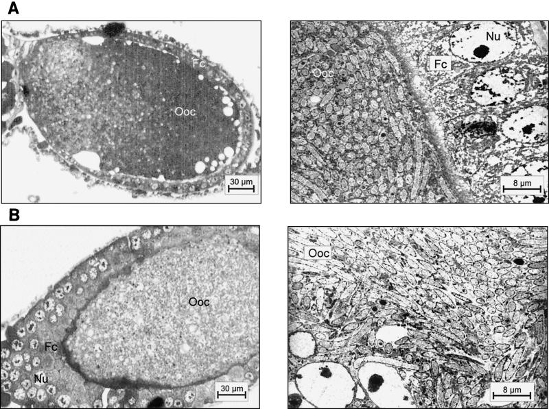
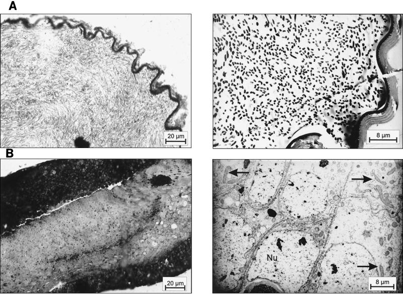
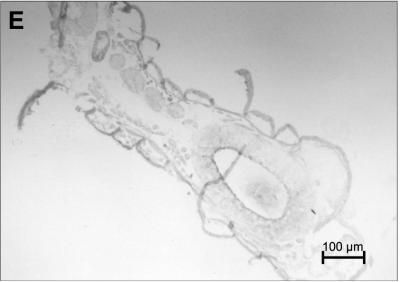
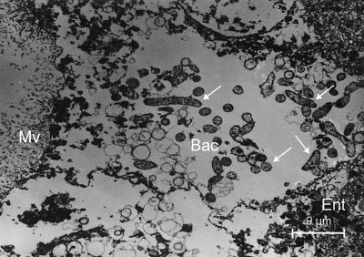
The distribution of endosymbiotic bacteria in different tissues of queens, males, and workers of the carpenter ant Camponotus floridanus was investigated by light and electron microscopy and by in situ hybridization. A large number of bacteria could be detected in bacteriocytes within the midguts of workers, young virgin queens, and males. Large amounts of bacteria were also found in the oocytes of workers and queens. In contrast, bacteria were not present in oocyte-associated cells or in the spermathecae of mature queens, although occasionally a small number of bacteria could be detected in the testis follicles of males. Interestingly, the number of bacteriocytes in mature queens was strongly reduced and the bacteriocytes contained only very few or no bacteria at all, although the endosymbionts were present in huge amounts in the ovaries of the same animals. During embryogenesis of the deposited egg, the bacteria were concentrated in a ring of endodermal tissue destined to become the midgut in later developmental stages. However, during larval development, bacteria could also be detected in other tissues although to a lesser extent. Only in the last-instar larvae were bacteria found exclusively in the midgut tissue within typical bacteriocytes. Tetracycline and rifampin efficiently cleansed C. floridanus workers of their symbionts and the bacteriocytes of these animals still remained empty several months after treatment had ceased. Despite the lack of their endosymbionts, these adult animals were able to survive without any obvious negative effect under normal cultivation conditions.
Figures

References
-
- Aksoy, S., X. Chen, and V. Hypsa. 1997. Phylogeny and potential transmission routes of midgut-associated endosymbionts of tsetse (Diptera: Glossinidae). Insect Mol. Biol. 6:183-190. - PubMed
-
- Baumann, P., L. Baumann, C. Y. Lai, D. Rouhbakhsh, N. A. Moran, and M. A. Clark. 1995. Genetics, physiology, and evolutionary relationships of the genus Buchnera: intracellular symbionts of aphids. Annu. Rev. Microbiol. 49:55-94. - PubMed
-
- Blochmann, F. 1882. Über das Vorkommen bakterienähnlicher Gebilde in den Geweben und Eiern verschiedener Insekten. Zentbl. Bakteriol. 11:234-240.
-
- Bolton, B. 1996. A new general catalogue of the ants of the world. Harvard University Press, Cambridge, Mass.
-
- Buchner, P. 1918. Vergleichende Eistudien. I. Die akzessorischen Kerne des Hymenoptereneies. Arch. Mikroskop. Anat. II 91:70-88.
Publication types
MeSH terms
Substances
LinkOut - more resources
Full Text Sources
