

Unveiling the organisms behind novel eukaryotic ribosomal DNA sequences from the ocean
- PMID: 12200313
- PMCID: PMC124113
- DOI: 10.1128/AEM.68.9.4554-4558.2002
Unveiling the organisms behind novel eukaryotic ribosomal DNA sequences from the ocean
Abstract
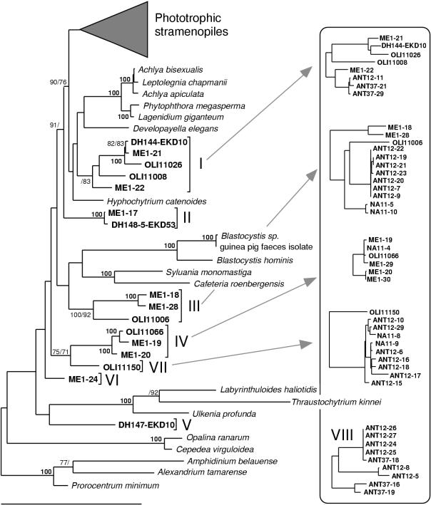
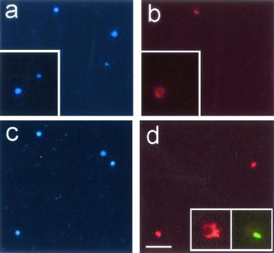
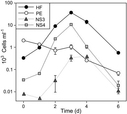
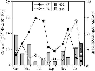
Despite the fact that the smallest eukaryotes (cells less than 5 micro m in diameter) play key roles in marine food webs, particularly in open oligotrophic areas, the study of their in situ diversity started just one year ago. Perhaps the most remarkable finding of the most recent studies has been the discovery of completely new phylogenetic lineages, such as novel clades belonging to the stramenopile and alveolate phyla. The two new groups account for a significant fraction of clones in genetic libraries from North Atlantic, equatorial Pacific, Antarctic, and Mediterranean Sea waters. However, the identities and ecological relevance of these organisms remain unknown. Here we investigate the phylogenetic relationships, morphology, in situ abundance, and ecological role of novel stramenopiles. They form at least eight independent clades within the stramenopile basal branches, indicating a large phylogenetic diversity within the group. Two lineages were visualized and enumerated in field samples and enrichments by fluorescent in situ hybridization using specific rRNA-targeted oligonucleotide probes. The targeted organisms were 2- to 3- micro m-diameter, round-shaped, nonpigmented flagellates. Further, they were found to be bacterivorous. One lineage accounted for up to 46% (average during an annual cycle, 19%) of heterotrophic flagellates in a coastal environment, providing evidence that novel stramenopiles are important and unrecognized components of the total stock of bacterial grazers.
Figures
Similar articles
-
Phylogenetic and ecological analysis of novel marine stramenopiles.Appl Environ Microbiol. 2004 Jun;70(6):3528-34. doi: 10.1128/AEM.70.6.3528-3534.2004. Appl Environ Microbiol. 2004. PMID: 15184153 Free PMC article.
-
Distribution and abundance of uncultured heterotrophic flagellates in the world oceans.Environ Microbiol. 2006 Sep;8(9):1515-22. doi: 10.1111/j.1462-2920.2006.01042.x. Environ Microbiol. 2006. PMID: 16913912
-
Study of genetic diversity of eukaryotic picoplankton in different oceanic regions by small-subunit rRNA gene cloning and sequencing.Appl Environ Microbiol. 2001 Jul;67(7):2932-41. doi: 10.1128/AEM.67.7.2932-2941.2001. Appl Environ Microbiol. 2001. PMID: 11425705 Free PMC article.
-
Unveiling new microbial eukaryotes in the surface ocean.Curr Opin Microbiol. 2008 Jun;11(3):213-8. doi: 10.1016/j.mib.2008.04.004. Epub 2008 Jun 13. Curr Opin Microbiol. 2008. PMID: 18556239 Review.
-
The diversity of small eukaryotic phytoplankton (< or =3 microm) in marine ecosystems.FEMS Microbiol Rev. 2008 Aug;32(5):795-820. doi: 10.1111/j.1574-6976.2008.00121.x. Epub 2008 Jun 28. FEMS Microbiol Rev. 2008. PMID: 18564290 Review.
Cited by
-
Picoeukaryotic plankton diversity at the Helgoland time series site as assessed by three molecular methods.Microb Ecol. 2006 Jul;52(1):53-71. doi: 10.1007/s00248-005-0062-x. Epub 2006 May 19. Microb Ecol. 2006. PMID: 16703447
-
Community structure and dynamics of small eukaryotes targeted by new oligonucleotide probes: new insight into the lacustrine microbial food web.Appl Environ Microbiol. 2009 Oct;75(19):6373-81. doi: 10.1128/AEM.00607-09. Epub 2009 Aug 7. Appl Environ Microbiol. 2009. PMID: 19666727 Free PMC article.
-
Defining DNA-based operational taxonomic units for microbial-eukaryote ecology.Appl Environ Microbiol. 2009 Sep;75(18):5797-808. doi: 10.1128/AEM.00298-09. Epub 2009 Jul 10. Appl Environ Microbiol. 2009. PMID: 19592529 Free PMC article.
-
Reverse evolution: driving forces behind the loss of acquired photosynthetic traits.PLoS One. 2009 Dec 29;4(12):e8465. doi: 10.1371/journal.pone.0008465. PLoS One. 2009. PMID: 20041159 Free PMC article.
-
Genetic diversity of microbial eukaryotes in anoxic sediment around fumaroles on a submarine caldera floor based on the small-subunit rDNA phylogeny.Extremophiles. 2005 Jun;9(3):185-96. doi: 10.1007/s00792-005-0432-9. Epub 2005 Mar 3. Extremophiles. 2005. PMID: 15744454
References
-
- Arndt, H. 2000. Functional diversity of heterotrophic flagellates in aquatic systems, p. 240-268. In B. S. C. Leadbeater and J. C. Green (ed.), The flagellates: unity, diversity and evolution. Taylor & Francis Press, London, United Kingdom.
-
- Bhattacharya, D., and L. Medlin. 1995. The phylogeny of plastids: a review based on comparisons of small-subunit ribosomal RNA coding regions. J. Phycol. 31:489-498.
-
- Caron, D. A., E. R. Peele, E. L. Lim, and M. R. Dennett. 1999. Picoplankton and nanoplankton and their trophic coupling in the surface waters of the Sargasso Sea south of Bermuda. Limnol. Oceanogr. 44:259-272.
Publication types
MeSH terms
Substances
LinkOut - more resources
Full Text Sources
Molecular Biology Databases
