. 2002 Sep;68(9):4623-8.
doi: 10.1128/AEM.68.9.4623-4628.2002.

Aquatic insects as a vector for Mycobacterium ulcerans

Affiliations

Aquatic insects as a vector for Mycobacterium ulcerans

Laurent Marsollier et al. Appl Environ Microbiol. 2002 Sep.

Abstract

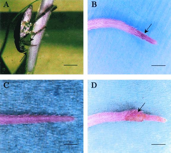
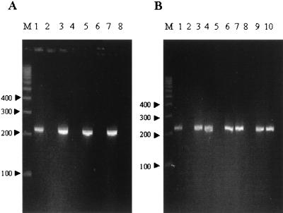
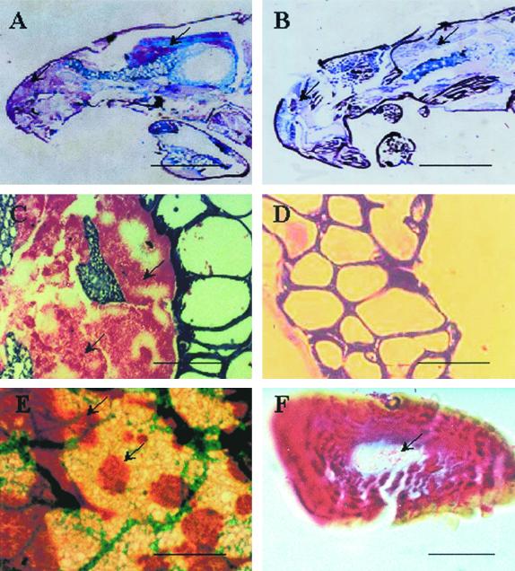
Mycobacterium ulcerans is an emerging environmental pathogen which causes chronic skin ulcers (i.e., Buruli ulcer) in otherwise healthy humans living in tropical countries, particularly those in Africa. In spite of epidemiological and PCR data linking M. ulcerans to water, the mode of transmission of this organism remains elusive. To determine the role of aquatic insects in the transmission of M. ulcerans, we have set up an experimental model with aquariums that mimic aquatic microenvironments. We report that M. ulcerans may be transmitted to laboratory mice by the bite of aquatic bugs (Naucoridae) that are infected with this organism. In addition, M. ulcerans appears to be localized exclusively within salivary glands of these insects, where it can both survive and multiply without causing any observable damage in the insect tissues. Subsequently, we isolated M. ulcerans from wild aquatic insects collected from a zone in the Daloa region of Ivory Coast where Buruli ulcer is endemic. Taken together, these results point to aquatic insects as a possible vector of M. ulcerans.


Figures

FIG. 1.
FIG. 1.
Transmission of M. ulcerans infection to BALB/c mice by biting insects (Naucoridae). (A) Water bug (N. cimicoides). Note its rostrum (lower arrow) and raptorial front legs (upper arrow), the femurs of which are greatly thickened, flattened, and adapted for grasping prey. A few minutes after a bite of the tail of a mouse by a Naucoris bug, a slight local erythema is noted (arrow) (B) which disappeared within 3 days (C). (D) After 57 days, active inflammatory lesions develop at the same site (arrow). This observation was made with six other mice. Scale bars: 0.5 cm (A) and 1 cm (B to D).
FIG. 2.
FIG. 2.
Identification of M. ulcerans based on detection of the IS2404 nested-PCR product. (A) Lanes contain a M. ulcerans DNA-positive control (lane 1); mock-specimen negative controls (lanes 2, 4, 6, and 8); and extractions from a water bug (Naucoridae) experimentally infected by M. ulcerans (strain 1G897) (lane 3), from skin biopsy material (day 57) from the tail of a BALB/c mouse bitten by an infected bug (lane 5), and from bacterial colonies isolated by culture from the salivary glands of a water bug (Naucoridae) (lane 7). (B) Nested-PCR amplification of DNAs extracted from M. ulcerans (lane 1) and salivary glands of Naucoridae captured in France that were experimentally infected by M. ulcerans (strain 1G897) (lane 3) and salivary glands of Naucoridae captured in Ivory Coast that were naturally infected by M. ulcerans (lane 4) or were not infected (lane 5). PCR was also performed on the two isolates (Nau.CI.001 and Nau.CI.002) cultured on Löwenstein-Jensen medium (lanes 6 and 7). Eight weeks after subcutaneous inoculation of BALB/c mouse tails with 104 bacilli (Nau.CI.001 and Nau.CI.002), PCR was performed on skin biopsy material from the mouse tails (lanes 9 and 10). Lanes 2 and 8 contain mock-specimen negative controls. Lane M contains molecular size markers (100-bp ladder; Gensura, San Diego, Calif.).
FIG. 3.
FIG. 3.
Localization of M. ulcerans in 5-μm-thick sections of tissues from infected water bugs. (A) Ziehl-Neelsen staining of sagittal sections of an experimentally infected bug of the order Naucoridae shows colonies of AFB restricted to the principal (left arrow) and accessory (right arrow) salivary glands. The latter are infiltrated by aggregates of AFB (C) and are immunostained with brown dye (arrows) and counterstained with light-green dye (E). No AFB were observed in tissues of noninfected insects (control group) (B) or in their salivary glands (D). The lumens of salivary canals in a silk (F) covering a raptorial leg show many AFB. Scale bars: 0.5 cm (A and B), 100 μm (C to E), and 25 μm (F).
FIG. 4.
FIG. 4.
Cutaneous lesions of a mouse tail due to M. ulcerans isolated from the salivary glands of a Naucoris bug captured in Ivory Coast. (A) BALB/c mouse tail showing an inflammatory ulceration (arrow) 8 weeks after subcutaneous inoculation of 104 bacilli (strain 01G897). (B and C) The same lesions (arrow) were observed in mouse tails inoculated with Nau.CI.001 (B) and Nau.CI.002 (C), which were isolated by culture from the salivary glands of water bugs captured in Ivory Coast. (D) Control mouse tail. Scale bars: 1.5 cm.

Similar articles

Cited by

References

    1. Asiedu, K., R. Sherpbier, and M. C. Raviglione. 2000. Buruli ulcer Mycobacterium ulcerans infection. WHO Global Buruli Ulcer initiative. Report 2000. World Health Organization, Geneva, Switzerland.
    1. Boom, R., C. J. A. Sol, M. M. M. Salimans, C. L. Jansen, P. M. E. Wertheim-van Dillen, and J. van der Noordaa. 1990. Rapid and simple method for purification of nucleic acids. J. Clin. Microbiol. 28:495-503. - PMC - PubMed
    1. Darie, H., T. Le Guyadec, and J. E. Touze. 1993. Epidemiological and clinical aspects of Buruli ulcer in Ivory Coast: 124 recent cases. Bull. Soc. Pathol. Exot. 86:272-276. (In French.) - PubMed
    1. De Gentile, P. L., C. Mahaza, F. Rolland, B. Carbonnelle, J. L. Verret, and D. Chabasse. 1992. Cutaneous ulcer from Mycobacterium ulcerans. Apropos of 1 case in French Guyana. Bull. Soc. Pathol. Exot. 85:212-214. (In French.) - PubMed
    1. Dobos, K. M., P. L. Small, M. Deslauriers, F. D. Quinn, and C. H. King. 2001. Mycobacterium ulcerans cytotoxicity in an adipose cell model. Infect. Immun. 69:7182-7186. - PMC - PubMed

Publication types