. 2002 Sep 1;30(17):3662-71.
doi: 10.1093/nar/gkf508.

Predicted structure and phyletic distribution of the RNA-binding protein Hfq

Affiliations

Predicted structure and phyletic distribution of the RNA-binding protein Hfq

Xueguang Sun et al. Nucleic Acids Res. .

Abstract

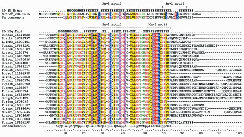
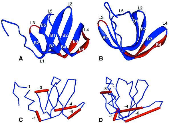
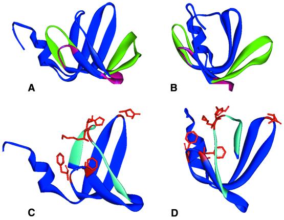
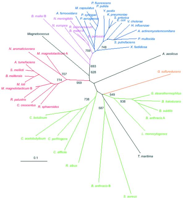
Hfq, a bacterial RNA-binding protein, was recently shown to contain the Sm1 motif, a characteristic of Sm and LSm proteins that function in RNA processing events in archaea and eukaryotes. In this report, comparative structural modeling was used to predict a three-dimensional structure of the Hfq core sequence. The predicted structure aligns with most major features of the Methanobacterium thermoautotrophicum LSm protein structure. Conserved residues in Hfq are positioned at the same structural locations responsible for subunit assembly and RNA interaction in Sm proteins. A highly conserved portion of Hfq assumes a structural fold similar to the Sm2 motif of Sm proteins. The evolution of the Hfq protein was explored by conducting a BLAST search of microbial genomes followed by phylogenetic analysis. Approximately half of the 140 complete or nearly complete genomes examined contain at least one gene coding for Hfq. The presence or absence of Hfq closely followed major bacterial clades. It is absent from high-level clades and present in the ancient Thermotogales-Aquificales clade and all proteobacteria except for those that have undergone major reduction in genome size. Residues at three positions in Hfq form signatures for the beta/gamma proteobacteria, alpha proteobacteria and low GC Gram-positive bacteria groups.


Figures

Figure 1
Figure 1
Multiple alignment of Hfq proteins from 26 bacterial genomes compared with the LSm protein from M.thermoautotrophicum and a consensus sequence for Sm proteins (shown above the alignment). Known secondary structure for the LSm protein and predicted structure for Hfq proteins are shown above the corresponding sequences: H, α helix; E, β strand. The Sm1 and Sm2 motifs of the Sm protein and the Sm1 and Xm2 motifs of Hfq proteins are shown. The 90% consensus shown below the alignment was derived using the following amino acid groupings. Positively charged residues (RKH) are shown as white letters on a red background; polar residues (p, KRHEDQNST) are shown as red letters; turn-like residues (t, ACDEGKNQRST) are green letters; bulky hydrophobic residues (h, ACLIVMHYFW) and the aliphatic subset of these type residues (l, LIVM) have a yellow background; aromatic residues (a, FHWY) are white letters with a purple background; small residues (s, ACDGNPSTV) are blue letters; tiny (u, AGS) are white letters with a blue background. Sequences are denoted by the species abbreviation followed by GI number. Species abbreviations: M.ther, M.thermoautotrophicum; B.halo, Bacillus halodurans; B.subt, Bacillus subtilis; L.inno, Listeria innocua; T.mari, Thermotoga maritima; C.acet, Clostridium acetobutylicum; A.caul, Azorhizobium caulinodans; C.cres, Caulobacter crescentus; M.loti, Mesorhizobium loti; B.meli, Brucella melitensis biovar Abortus; S.meli, Sinorhizobium meliloti; P.mult, Pasteurella multocida; P.prof, Photobacterium profundum; H.infl, Haemophilus influenzae; V.chol, Vibrio cholerae; Y.pest, Yersinia pestis; Y.ente, Yersinia enterocolitica; P.caro, Pectobacterium carotovorum; E.coli, E.coli; S.typh, Salmonella typhimurium; S.flex, Shigella flexneri; P.aeru, P.aeruginosa; X.fast, Xylella fastidiosa; N.meni, Neisseria meningitidis; A.aeol, Aquifex aeolicus; B.anth, Bacillus anthracis; S.aure, S.aureus.
Figure 2
Figure 2
(A and B) Ribbon representations of two views of the crystal structure of an archaeal Sm protein (PDB accession number 1i81) rotated by 90Ε. Images are produced by RasMol program (http://www.bernstein-plus-sons.com/software/RasMol_2.7.1). (C and D) 3D line representations of the Hfq structure predicted by the 3D-PSSM web server using the above archaeal Sm protein as template. The views shown in (C) and (D) are the same as in (A) and (B) respectively. The locations of Hfq residues that are inserted or deleted when compared with the template are represented by thin and thick bars respectively, and accompanied by numbers indicating the number of residues involved. Labels B1–B5 correspond to the β strands β1–β5; labels L1–L5 correspond to the loops.
Figure 3
Figure 3
The 3D structure of Hfq generated by SWISS-MODEL program using the same archaeal LSmα protein determined to be the best template by 3D PSSM. (A and B) Front and side views of the predicted Hfq structure as well as the template Sm structure. The backbone features that are constant in both structures are illustrated in blue. Differing structural elements are shown by using a red ribbon for Hfq and a green ribbon to illustrate the LSmα protein backbone. The aqua ribbon illustrates the β4 strand residues SQMVY and β5 strand residues AISTVV. (C and D) Front and side views of the predicted Hfq model with several potential RNA-interacting residues shown in stick model representation: Lys31 in loop 2, Phe39 and Phe42 in loop 3, Lys56 and His57 in loop 5.
Figure 4
Figure 4
Molecular representation of the β4 and β5 strands in Hfq model (A) and LSmα protein (B). Overlapping representation of both is shown in (C).
Figure 5
Figure 5
Unrooted neighbor-joining tree inferred by analysis of Hfq protein sequences. Sequences were aligned using CLUSTAL program and all positions with gaps were excluded from the analysis. Bootstrap values of >600 are displayed at deep nodes only. Color code: green, low GC Gram-positive bacteria; red, alpha proteobacteria; purple, beta proteobacteria; blue, gamma proteobacteria; orange, delta proteobacteria. Aquifecales-Thermatogales and unclassified Magnetococcus are shown in black.
Figure 6
Figure 6
Conserved amino acid residues specific to Hfq proteins from major bacterial groups defined by phylogenetic analysis. Multiple alignment of Hfq sequences is subdivided according to bacterial groups inferred from the tree shown in Figure 5. Positions where amino acid conservation are group specific are shown.

References

    1. Kajitani M. and Ishihama,A. (1991) Identification and sequence determination of the host factor gene for bacteriophage Q beta. Nucleic Acids Res., 19, 1063–1066. - PMC - PubMed
    1. Franze de Fernandez M.T., Eoyang,L. and August,J.T. (1968) Factor fraction required for the synthesis of bacteriophage Q beta-RNA. Nature, 219, 588–590. - PubMed
    1. Carmichael G.G., Weber,K., Niveleau,A. and Wahba,A.J. (1975) The host factor required for RNA phage Qbeta RNA replication in vitro. Intracellular location, quantitation, and purification by polyadenylate-cellulose chromatography. J. Biol. Chem., 250, 3607–3612. - PubMed
    1. Kajitani M., Kato,A., Wada,A., Inokuchi,Y. and Ishihama,A. (1994) Regulation of the Escherichia coli hfq gene encoding the host factor for phage Q beta. J. Bacteriol., 176, 531–534. - PMC - PubMed
    1. Azam T.A, Hiraga,S. and Ishihama,A. (2000) Two types of localization of the DNA-binding proteins within the Escherichia coli nucleoid. Genes Cells, 5, 613–626. - PubMed

Publication types

MeSH terms