. 2002 Sep;39(3):229-36.
doi: 10.1002/glia.10102.

Remyelination of the spinal cord following intravenous delivery of bone marrow cells

Affiliations

Remyelination of the spinal cord following intravenous delivery of bone marrow cells

Yukinori Akiyama et al. Glia. 2002 Sep.

Abstract

Bone marrow contains a population of pluripotent cells that can differentiate into a variety of cell lineages, including neural cells. When injected directly into the demyelinated spinal cord they can elicit remyelination. Recent work has shown that following systemic delivery of bone marrow cells functional improvement occurs in contusive spinal cord injury and stroke models in rat. We report here that secondary to intravenous introduction of an acutely isolated bone marrow cell fraction (mononuclear fraction) from adult rat femoral bones separated on a density gradient, ultrastructurally defined remyelination occurs throughout a focal demyelinated spinal cord lesion. The anatomical pattern of remyelination was characteristic of both oligodendrocyte and Schwann cell myelination; conduction velocity improved in the remyelinated axons. When the injected bone marrow cells were transfected to express LacZ, beta-galactosidase reaction product was observed in some myelin-forming cells in the spinal cord. Intravenous injection of other myelin-forming cells (Schwann cells and olfactory ensheathing cells) or the residual cell fraction of the gradient did not result in remyelination, suggesting that remyelination was specific to the delivery of the mononuclear fraction. While the precise mechanism of the repair, myelination by the bone marrow cells or facilitation of an endogenous repair process, cannot be fully determined, the results demonstrate an unprecedented level of myelin repair by systemic delivery of the mononuclear cells.


Figures

Fig. 1
Fig. 1
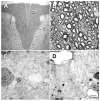
Morphology of dorsal funiculus from control, demyelinated, and sham cell transplanted rat spinal cord. Normal dorsal funiculus (arrowheads) of the spinal cord is shown in A and B at low and high power, respectively. C: High-power field of demyelinated axons in control lesion with no cell injections. No myelination was observed following intravenous delivery of Schwann cells (D). DF, dorsal funiculus; DH, dorsal horn. Scale bar, 100 μm in A; 10 μm in B–D.
Fig. 2
Fig. 2
Dorsal funiculus following intravenous delivery of mononuclear cells. (A) Low power micrograph of dorsal funiculus lesion zone three weeks after intravenous bone marrow delivery. Remyelinated axons in the center (B) and lateral edge (C) of the lesion. Remyelination was observed throughout the lesion zone. However, some areas had various degrees of remyelination. This is shown in D, where an area of intense myelination (left) is adjacent to an area with promyelinating profiles. Scale bar, 100 μm in A; 15 μm in B and C; 6 μm in D.
Fig. 3
Fig. 3
Electron micrographs of remyelinated axons in the dorsal funiculus of the spinal cord after bone marrow transplantation. A: Normal dorsal funiculus axons. B: A field of axons showing some with myelin and others without. Some myelin-forming cells were similar to peripheral myelin-forming cells, characterized by large cytoplasmic and nuclear domains (C) and a basement membrane (arrowheads in inset). D: Other myelinated axons did not have the Schwann cell-like morphology and were similar to axons remyelinated by oligodendrocytes. Scale bar, 5 μm in A and B; 1.5 μm in C; 3 μm in D.
Fig. 4
Fig. 4
A: Two mm whole mount of coronally cut spinal cord from a rat that was injected intravenously with LacZ-transfected rat bone marrow cells. Note the intense blue β-galactosidase reaction in the lesion zone only. Numerous cells with β-galactosidase blue reaction product are observed in the remyelinated dorsal funiculus 3 weeks after intravenous delivery of LacZ-transfected rat bone marrow cells (B). C: Higher-power photomicrograph showing a Schwann-like cell with blue β-galactosidase reaction product in the cytoplasm. β-galactosidase reaction product is seen in cell bodies reminiscent of oligodendrocytes (D). E: Micrograph of normal white matter from same animal as that for B–D. Note the paucity of β-galactosidase reaction product in the nonlesion white matter. The sections in B–E were counterstained with 0.15% basic fuchsin. F: Micrograph showing PKH26 fluorescence in the dorsal funiculus lesion zone after intravenous delivery of labeled bone marrow cells. Dashed line indicates dorsal funiculus. Scale bar, 1 mm in A and F; 8 μm in B; 4 μm in C; 6 μm in D; 30 μm in E.
Fig. 5
Fig. 5
A: Compound action potentials recorded at 1.0 mm increments longitudinally along the dorsal columns in normal, demyelinated (EB-X lesion) and after bone marrow cells were delivered intravenously in an EB-X lesioned rat. Note that multicomponent waves were observed in the spinal cord from the bone marrow-injected rat. The dashed lines link the peak negativities of three primary negativities. Histograms of conduction velocity (error bars indicate SEM) of dorsal column axons obtained from normal, demyelinated, and the fastest component (dashed line with 1) of recordings obtained from the bone marrow injected rats are shown in B. At 36°C, conduction velocity of the early negativity in control, demyelinated, and bone marrow delivered groups were 10.88 ± 1.21 m/s (n = 15), 0.83 ± 0.05 m/s (n = 15), and 6.21 ± 0.82 m/s (n = 15), respectively. Double asterisk: P < 0.001, control vs. demyelinated; asterisk: P < 0.01, demyelinated vs. transplanted.

References

    1. Akiyama Y, Honmou O, Kato T, Uede T, Hashi K, Kocsis JD. Transplantation of clonal neural precursor cells derived from adult human brain establishes functional peripheral myelin in the rat spinal cord. Exp Neurol. 2001;167:27–39. - PubMed
    1. Azizi SA, Stokes D, Augelli BJ, Digirolamo C, Prockop DJ. Engraftment and migration of human bone marrow stromal cells implanted in the brains of albino rats: similarities to astrocyte grafts. Proc Natl Acad Sci USA. 1998;95:3908–3913. - PMC - PubMed
    1. Bjornson CR, Rietze RL, Reynolds BA, Magli MC, Vescovi AL. Turning brain into blood: a hematopoietic fate adopted by adult neural stem cells in vivo. Science. 1999;283:534–537. - PubMed
    1. Blakemore WF. Limited remyelination of CNS axons by Schwann cells transplanted into the sub-arachnoid space. J Neurol Sci. 1984;64:265–276. - PubMed
    1. Blakemore WF, Crang AJ. The use of cultured autologous Schwann cells to remyelinate areas of persistent demyelination in the central nervous system. J Neurol Sci. 1985;70:207–223. - PubMed

Publication types

LinkOut - more resources