. 2002;3(1):22.
doi: 10.1186/rr172. Epub 2002 Jul 10.

Mechanism of cigarette smoke condensate-induced acute inflammatory response in human bronchial epithelial cells

Affiliations

Mechanism of cigarette smoke condensate-induced acute inflammatory response in human bronchial epithelial cells

Gary R Hellermann et al. Respir Res. 2002.

Abstract

Background: To demonstrate the involvement of tobacco smoking in the pathophysiology of lung disease, the responses of pulmonary epithelial cells to cigarette smoke condensate (CSC) - the particulate fraction of tobacco smoke - were examined.

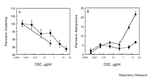
Methods: The human alveolar epithelial cell line A549 and normal human bronchial epithelial cells (NHBEs) were exposed to 0.4 microg/ml CSC, a concentration that resulted in >90% cell survival and <5% apoptosis. Changes in gene expression and signaling responses were determined by RT-PCR, western blotting and immunocytofluorescence.

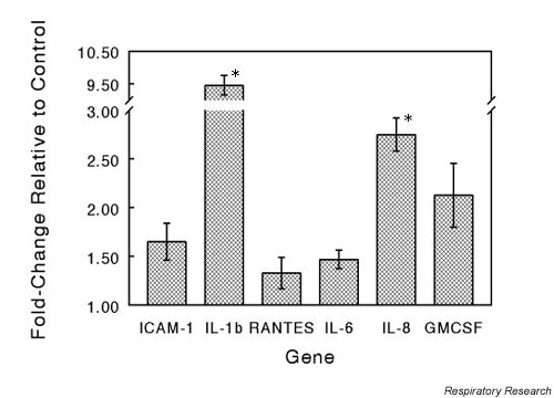
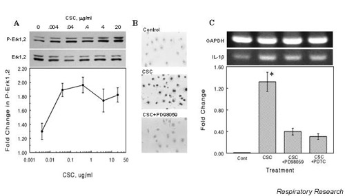
Results: NHBEs exposed to CSC showed increased expression of the inflammatory mediators sICAM-1, IL-1beta, IL-8 and GM-CSF, as determined by RT-PCR. CSC-induced IL-1beta expression was reduced by PD98059, a blocker of mitogen-actived protein kinase (MAPK) kinase (MEK), and by PDTC, a NFkappaB inhibitor. Analysis of intracellular signaling pathways, using antibodies specific for phosphorylated MAPKs (extracellular signal-regulated kinase [ERK]-1/2), demonstrated an increased level of phosphorylated ERK1/2 with increasing CSC concentration. Nuclear localization of phosphorylated ERK1/2 was seen within 30 min of CSC exposure and was inhibited by PD98059. Increased phosphorylation and nuclear translocation of IkappaB was also seen after CSC exposure. A549 cells transfected with a luciferase reporter plasmid containing a NFkappaB-inducible promoter sequence and exposed to CSC (0.4 microg/ml) or TNF-alpha (50 ng/ml) had an increased reporter activity of approximately 2-fold for CSC and 3.5-fold for TNF-alpha relative to untreated controls.

Conclusion: The acute phase response of NHBEs to cigarette smoke involves activation of both MAPK and NFkappaB.


Figures

Figure 1
Figure 1
Effect of CSC on viability and apoptosis in A549 cells and NHBEs. Cells were grown to about 70% confluence in 12-well plates (Panel A, viability) or 8-well chamber slides (Panel B, apoptosis). Cells were exposed to the indicated concentration of CSC for 30 min then rinsed with PBS and processed for each assay. Viability was determined by trypan blue vital staining and expressed as percent of total cells. Apoptosis was measured by fluorescent TUNEL assay and is expressed as the percent of cells exhibiting apoptosis relative to the total number of cells. The values are the mean ± SEM for n = 3. (--●-- A549; --▲-- NHBE) CSC = cigarette smoke condensate; NBHE = normal human bronchial epithelial cell; PBS = phosphate-buffered saline; SEM = standard error of the mean; TUNEL = deoxynucleotidyl transferase-mediated dUTP nick end labeling
Figure 2
Figure 2
Effect of CSC treatment on proinflammatory gene expression. NHBEs were exposed to CSC at a concentration of 0.4 μg/ml for 30 min at 37°C. Total RNA was isolated and specific gene expression measured by RT-PCR assay. Target gene expression levels were normalized to the level of GAPDH expression in each reaction and expressed as fold change relative to control. The values are means ± SEM for n = 3. Significant differences at P < 0.05 are indicated by *. CSC = cigarette smoke condensate; NBHE = normal human bronchial epithelial cell; RT-PCR = reverse transcriptase-polymerase chain reaction; SEM = standard error of the mean
Figure 3
Figure 3
CSC-induced phosphorylation of ERK1/2 and IL-1β expression. (A) CSC induces ERK1/2 phosphorylation. NHBEs were incubated with the indicated concentrations of CSC for 30 min at 37°C. Whole-cell protein extracts were prepared from cell lysates, aliquots of 10 μg of protein were separated by SDS-PAGE, transferred to nitrocellulose and probed with an antibody to phosphorylated ERK1/2. Blots were then stripped and reprobed for ERK1/2. The band intensities were determined by densitometry and the fold-change relative to control was plotted as means ± SEM. (B) Nuclear localization of phosphorylated ERK1/2 in NHBEs. Cells were incubated with 0.4 μg/ml CSC for 30 min at 37°C, then fixed with paraformaldehyde and probed with antibody to phosphorylated ERK1/2. Visualization was achieved by incubating the cells with HRP-conjugated secondary antibody and bisbenzamidine substrate. Cells were treated with vehicle alone (Control), with CSC (CSC), or with CSC and the MEK inhibitor, PD98059 (20 μM) (CSC + PD98059). (C) Inhibition of ERK1/2 or IκB phosphorylation attenuates IL-1β expression in A549. Cells were treated with 50 μg/ml PD98059 or 1 mM pyrrolidine dithiocarbamate (PDTC) for 30 min then exposed to 0.4 μg/ml CSC for 60 min. RT-PCR was performed on RNA extracted from these cells using IL-1β-specific primers. Band densities were normalized to GAPDH levels, expressed as fold change relative to untreated controls, and plotted as mean ± SEM for n = 3. The * indicates significance at P < 0.05. CSC = cigarette smoke condensate; ERK = extracellular signal-regulated kinase; IκB = inhibitor of NFκB; MEK = MAPK kinase; NBHE = normal human bronchial epithelial cell.
Figure 4
Figure 4
Phosphorylation of IκB and expression of NFκB-luciferase reporter gene in cells exposed to CSC. (A) CSC exposure results in phosphorylation of IκB in NHBEs. Cells were grown to approximately 80% confluency then treated with the indicated concentrations of CSC for 30 min at 37°C. Whole-cell protein extracts were prepared and 10 μg aliquots were separated by SDS-PAGE, blotted and probed with an antibody to phosphorylated IκB. The blot was then stripped and reprobed for IκB. The band intensities for phosphorylated IκB were determined by densitometric scanning and normalized to the levels of IκB in each lane. Values are means ± SEM for n = 3. (B) CSC-induced immunofluorescent staining for phosphorylated IκB in NHBEs. Cells were treated with vehicle (Control) or 0.04 μg/ml CSC (CSC) for 30 min at 37°C. Cells were fixed with paraformaldehyde and probed with antibody to phosphorylated IκB. (C) and (D) Effect of CSC on expression of an NFκB-regulated luciferase reporter gene in A549 cells. Cells were transfected with a plasmid containing an NFκB binding site upstream of a luciferase reporter sequence. The transfected cells were exposed to 0.4 μg/ml CSC for 30 min at 37°C (C) or increasing doses of CSC (D) and lysates were assayed for luciferase activity using a luminometer. Differences in transfection efficiency were normalized by cotransfection with a LacZ-containing plasmid. The treatments were performed in triplicate and expressed as mean ± SEM for n = 3. CSC = cigarette smoke condensate; IκB = inhibitor of NFκB; SEM = standard error of the mean; NFκB = nuclear factor-kappa B; NHBE = normal human bronchial epithelial cell.

References

    1. Office of Environmental Health Hazard Assessment: Health Effects of Exposure to Environmental Tobacco Smoke. California Environmental Protection Agency; 1997.
    1. Floreani AA, Rennard SI. The role of cigarette smoke in the pathogenesis of asthma and as a trigger for acute symptoms. Curr Opin Pulm Med. 1999;5:38–46. doi: 10.1097/00063198-199901000-00007. - DOI - PubMed
    1. Adler KB, Fischer BM, Wright DT, Cohen LA, Becker S. Interactions between respiratory epithelial cells and cytokines: relationships to lung inflammation. Ann NY Acad Sci. 1994;725:128–145. - PubMed
    1. Rusznak C, Sapsford RJ, Devalia JL, Shah SS, Hewitt EL, Lamont AG, Davies RJ, Lozewicz S. Interaction of cigarette smoke and house dust mite allergens on inflammatory mediator release from primary cultures of human bronchial epithelial cells. Clin Exp Allergy. 2001;31:226–38. doi: 10.1046/j.1365-2222.2001.01000.x. - DOI - PubMed
    1. Mio T, Romberger DJ, Thompson AB, Robbins RA, Heires A, Rennard SI. Cigarette smoke induces interleukin-8 release from human bronchial epithelial cells. Am J Respir Crit Care Med. 1997;155:1770–6. - PubMed

Publication types

MeSH terms

LinkOut - more resources