. 2002 Sep 1;543(Pt 2):413-24.
doi: 10.1113/jphysiol.2002.022020.

An external site controls closing of the epithelial Na+ channel ENaC

Affiliations

An external site controls closing of the epithelial Na+ channel ENaC

Stephan Kellenberger et al. J Physiol. .

Abstract

Members of the ENaC/degenerin family of ion channels include the epithelial sodium channel (ENaC), acid-sensing ion channels (ASICs) and the nematode Caenorhabditis elegans degenerins. These channels are activated by a variety of stimuli such as ligands (ASICs) and mechanical forces (degenerins), or otherwise are constitutively active (ENaC). Despite their functional heterogeneity, these channels might share common basic mechanisms for gating. Mutations of a conserved residue in the extracellular loop, namely the 'degenerin site' activate all members of the ENaC/degenerin family. Chemical modification of a cysteine introduced in the degenerin site of rat ENaC (betaS518C) by the sulfhydryl reagents MTSET or MTSEA, results in a approximately 3-fold increase in the open probability. This effect is due to an 8-fold shortening of channel closed times and an increase in the number of long openings. In contrast to the intracellular gating domain in the N-terminus which is critical for channel opening, the intact extracellular degenerin site is necessary for normal channel closing, as illustrated by our observation that modification of betaS518C destabilises the channel closed state. The modification by the sulfhydryl reagents is state- and size-dependent consistent with a conformational change of the degenerin site during channel opening and closing. We propose that the intracellular and extracellular modulatory sites act on a common channel gate and control the activity of ENaC at the cell surface.


Figures

Figure 1
Figure 1. Hypothetical model of the ENaC pore
The cross-section shows the extracellular pore opening that narrows down to the amiloride binding site and the selectivity filter (where a Na+ ion is shown) and from there opens towards the transmembrane region and the cytoplasmic side. Experimental evidence indicates that the pre-M2 segments of all three subunits form the extracellular pore entry. Shown are the pre-M2 segments of two α subunits (left and right) and the β subunit in the back. The γ subunit located on the side of the viewer is not shown. The DEG residue is indicated for the α subunits (αS576). Amiloride binds to αS583 and the corresponding Gly residues in the β and γ subunit. The vestibule narrows down to the selectivity filter formed by αG587, βG529 and γS541 residues and the ring of Ser residues (αS589 and analogues). The N-terminal gating domain HG is indicated on the cytoplasmic side of the protein. S, G & H represent the amino acids Ser, Gly and His, respectively.
Figure 2
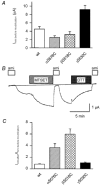
Figure 2. Effects of chemical modification on macroscopic currents
Whole-cell amiloride-sensitive currents (Iami). A, current expression of WT ENaC and the DEG mutants listed. B, trace of an experiment with an oocyte expressing the βS518C mutant. Extracellular application of reagents and drugs is indicated by bars. ami, 10 μM amiloride; MTSET, 1 mm MTSET; DTT, 10 mm dithiothreitol. C, increase in amiloride-sensitive current after 5 min incubation in 1 mm MTSET.
Figure 3
Figure 3. State-dependent modification of βS518C by MTSET
A, trace of an outside-out patch containing two active βS518C channels. Amiloride (10 μM) was first removed from the extracellular solution to allow normal channel opening and the sulfhydryl reagent MTSET (2 mm) was added at the time indicated. During the solution changes, small artefacts in the trace due to the solution change are visible. The arrows point to modification events. Holding voltage −100 mV. B, reversal of sulfhydryl modification of βS518C by DDT. Channels had previously been modified by MTSET. Channel activity appears after switching to the amiloride-free solution. Unitary current amplitude at −80 mV in this experiment was 0.80 pA (unmodified), 0.67 pA (MTSET) and 0.78 pA (DTT). C, outside-out patch containing one βS518C channel that showed only rare, extremely short openings during the time of MTSET application. Modification occurred during the first opening of longer duration (arrow). D, the inverse of the duration of MTSET application until successful modification is plotted versus the open probability (Po) of the channel during this time.
Figure 4
Figure 4. Modification of βS518C by MTSEA
Outside-out patches containing βS518C at holding voltage of −100 mV. Modification occurred while the channel was closed (upper trace) or while it was open (lower trace).
Figure 5
Figure 5. Effect of modification on βS518C single-channel kinetics
A, changes in single-channel open probability (Po) due to modification by MTSET or MTSEA. Po before and after modification by MTSEA (left panel) and MTSET (right panel). Po was determined during the minute prior to and after, modification, respectively. The Po data are from patches that contained single active channels. Holding potential was −100 mV. Filled symbols represent individual experiments. B, open and closed time distributions of βS518C channels before and after modification. Time distributions were obtained from single-channel excised outside-out patches at −100 mV. The time constants and the relative weights of the components are listed in Table 1. The overall Po of the data used for the kinetic analysis was 0.52 (unmodified), 0.95 (MTSEA-modified) and 0.97 (MTSET-modified). The number of exponential components of the fit was determined according to Colquhoun & Sigworth (1995) by visual inspection of the histograms and by the criterion of whether a given number of components was required to fit histograms derived from individual experiments. Therefore, open times of unmodified and MTSET-modified channels were fitted with two components. The open time distribution of MTSEA-modified channels was then fitted with two components to be better able to compare these data with those obtained in the other two conditions. Closed times were fitted with two exponential components.
Figure 6
Figure 6. Dependence of channel gating on the intracellular HG motif and the DEG site
A, outside-out patch containing several αH94A ENaC channels at a holding voltage of −100 mV. B, increase in amiloride-sensitive whole-cell currents due to modification of channels co-expressing βS518C with mutations in the pre-M1 gating domain. Oocytes were incubated for 5 min in 1 mm MTSET. The relative increase in INa was not significantly different between the single and the double mutants (P < 0.05). Initial whole-cell currents of the mutants were 3.8 ± 2.1 μA (βS518C), 0.06 ± 0.02 μA (αH94AβS518C), and 0.14 ± 0.13 μA (αG95SβS518C). C, representative traces from an outside-out patch containing αH94AβS518C after modification by MTSET, at a holding voltage of −100 mV. D, dwell time distributions of αH94AβS518C channels after modification by MTSET. Time distributions were obtained from single-channel excised outside-out patches at −100 mV. The time constants and the relative weights of the components are listed in Table 1.
Figure 7
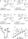
Figure 7. Possible involvement of DEG residues in pore functions
A-D, single-channel current-voltage relation from outside-out patches (n = 3–5 patches per condition). A, Li+ current of βS518C before (○) and after (▪) modification by MTSET (unitary conductance was 7.8 ± 0.9 pS (unmodified) and 6.8 ± 0.5 pS (modified) in direct comparison). B, Na+ current of βS518C before (○) and after (▪) modification by MTSET (4.4 ± 0.2/4.1 ± 0.3 pS). C, Li+ current of βS518C before (○) and after (▪) modification by MTSEA (9.9 ± 1.2/5.6 ± 0.7 pS). D, Li+ current of αS576C before (○) and after (▪) modification by MTSET (6.6 ± 0.6/7.1 ± 1.2 pS). E and F, two-electrode voltage-clamp recordings from oocytes expressing the βS518C mutant. E, current trace at holding voltage of −100 mV, the times of addition of amiloride (10 μM) or Cd2+ (10 mm) are indicated. F, current-voltage relationship of the macroscopic amiloride-sensitive current, Iami norm. in the absence (○) and presence (▪) of 6 mm Cd2+ (n = 4).
Figure 8
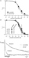
Figure 8. Amiloride block before and after chemical modification of DEG residues
Inhibition curves of Na+ current (Irelative)carried by WT (A) and the individual DEG mutants (B) before (filled symbols) and after modification by MTS-PtREA (open symbols). Continuous lines are fits to the concentration-dependence of inhibition. The inhibition curves were obtained using two-electrode voltage clamp at −100 mV. C, washout of amiloride and benzamil from βS518 channels measured in excised outside-out macropatches at −100 mV. The solution change is indicated by the arrow. The speed of the solution change was tested by changing from K+ to Li+ solution (black trace). Amiloride and benzamil washout was measured before (dark grey traces) and after (light grey traces) modification by MTSET. The maximal currents were normalised for better comparison of the time course. Exponential fits to this and similar experiments determined time constants of current increase of 0.58 ± 0.05 and 0.50 ± 0.15 s due to amiloride washout before and after MTSET incubation, respectively, and the corresponding values were 10.8 ± 4.4 s and 8.7 ± 0.7 s for benzamil washout (n = 2–3 each), respectively.

Similar articles

Cited by

References

    1. Adams CM, Anderson MG, Motto DG, Price MP, Johnson WA, Welsh MJ. Ripped pocket and pickpocket, novel Drosophila Deg/Enac subunits expressed in early development and in mechanosensory neurons. Journal of Cell Biology. 1998a;140:143–152. - PMC - PubMed
    1. Adams CM, Snyder PM, Price MP, Welsh MJ. Protons activate brain Na+ channel 1 by inducing a conformational change that exposes a residue associated with neurodegeneration. Journal of Biological Chemistry. 1998b;273:30204–30207. - PubMed
    1. Adams CM, Snyder PM, Welsh MJ. Paradoxical stimulation of a DEG/ENaC channel by amiloride. Journal of Biological Chemistry. 1999;274:15500–15504. - PubMed
    1. Champigny G, Voilley N, Waldmann R, Lazdunski M. Mutations causing neurodegeneration in Caenorhabditis elegans drastically alter the pH sensitivity and inactivation of the mammalian H+-gated Na+ channel MDEG1. Journal of Biological Chemistry. 1998;273:15418–15422. - PubMed
    1. Chraibi A, Vallet V, Firsov D, Hess SK, Horisberger JD. Protease modulation of the activity of the epithelial sodium channel expressed in Xenopus oocytes. Journal of General Physiology. 1998;111:127–138. - PMC - PubMed

Publication types

LinkOut - more resources