

Topical applications of caffeine or (-)-epigallocatechin gallate (EGCG) inhibit carcinogenesis and selectively increase apoptosis in UVB-induced skin tumors in mice
- PMID: 12205293
- PMCID: PMC129466
- DOI: 10.1073/pnas.182429899
Topical applications of caffeine or (-)-epigallocatechin gallate (EGCG) inhibit carcinogenesis and selectively increase apoptosis in UVB-induced skin tumors in mice
Abstract
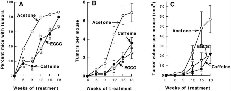
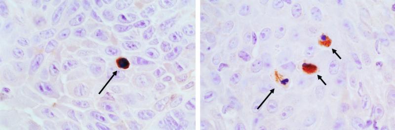
SKH-1 hairless mice were irradiated with ultraviolet B (UVB) twice weekly for 20 weeks. These tumor-free mice, which had a high risk of developing skin tumors during the next several months, were then treated topically with caffeine (6.2 micromol) or (-)-epigallocatechin gallate (EGCG; 6.5 micromol) once a day 5 days a week for 18 weeks in the absence of further treatment with UVB. Topical applications of caffeine to these mice decreased the number of nonmalignant and malignant skin tumors per mouse by 44% and 72%, respectively. Topical applications of EGCG decreased the number of nonmalignant and malignant tumors per mouse by 55% and 66%, respectively. Immunohistochemical analysis showed that topical applications of caffeine or EGCG increased apoptosis as measured by the number of caspase 3-positive cells in nonmalignant skin tumors by 87% or 72%, respectively, and in squamous cell carcinomas by 92% or 56%, respectively, but there was no effect on apoptosis in nontumor areas of the epidermis. Topical applications of caffeine or EGCG had a small inhibitory effect on proliferation in nonmalignant tumors as measured by BrdUrd labeling (16-22%), and there was also a similar, but nonsignificant, inhibitory effect on proliferation in malignant tumors. The results suggest a need for further studies to determine whether topical applications of caffeine or EGCG can inhibit sunlight-induced skin cancer in humans.
Figures
References
-
- Nataraj A. J., Trent, J. C., II & Ananthaswamy, H. N. (1995) Photochem. Photobiol. 62, 218-230. - PubMed
-
- McKenzie R., Connor, B. & Bodeker, G. (1999) Science 285, 1709-1711. - PubMed
-
- Stern R. S., Weinstein, M. C. & Baker, S. G. (1986) Arch. Dermatol. 122, 537-545. - PubMed
-
- Glanz K., Saraiya, M. & Wechsler, H. (2002) Morbid. Mortal. Wkly. Rep. 51, 1-18. - PubMed
-
- Lou Y.-R., Lu, Y.-P., Xie, J.-G., Huang, M.-T. & Conney, A. H. (1999) Nutr. Cancer 33, 146-153. - PubMed
Publication types
MeSH terms
Substances
Grants and funding
LinkOut - more resources
Full Text Sources
Other Literature Sources
Medical
Molecular Biology Databases
Research Materials
