. 2002 Nov;71(5):1161-7.
doi: 10.1086/344395. Epub 2002 Aug 29.

A gene mutated in nephronophthisis and retinitis pigmentosa encodes a novel protein, nephroretinin, conserved in evolution

Affiliations

A gene mutated in nephronophthisis and retinitis pigmentosa encodes a novel protein, nephroretinin, conserved in evolution

Edgar Otto et al. Am J Hum Genet. 2002 Nov.

Abstract

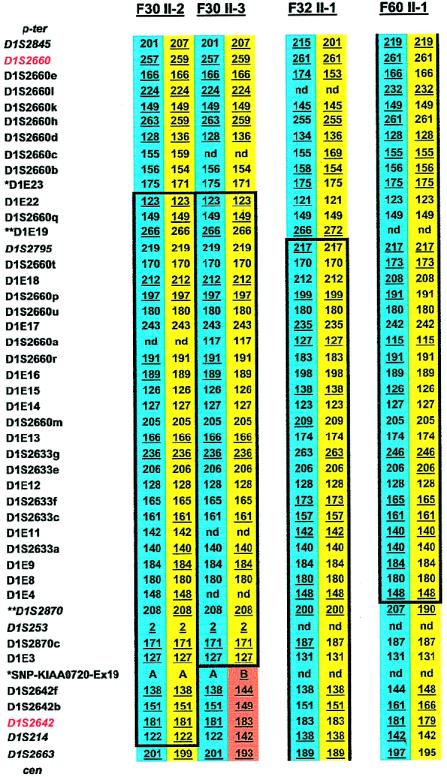
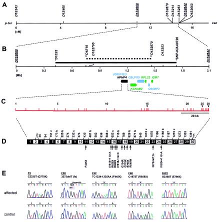
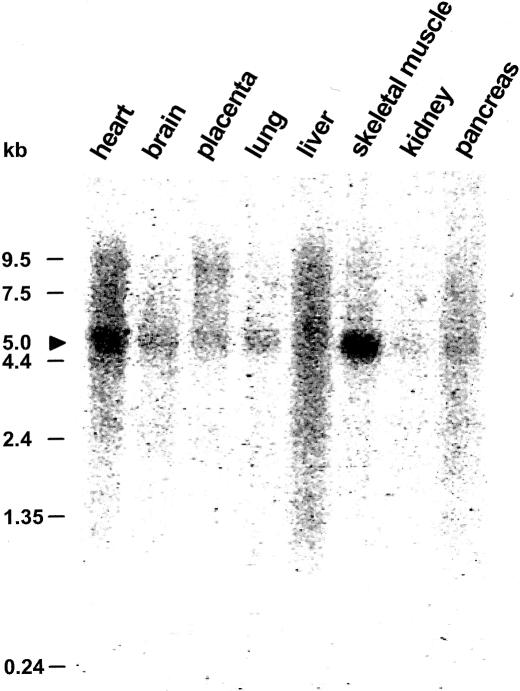
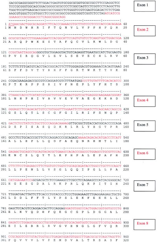
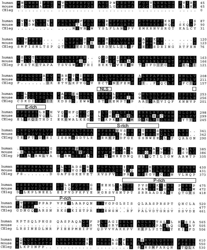
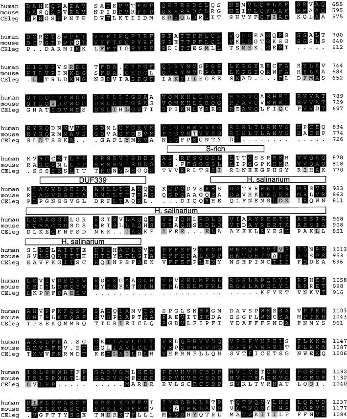
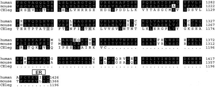
Nephronophthisis (NPHP) comprises a group of autosomal recessive cystic kidney diseases, which constitute the most frequent genetic cause for end-stage renal failure in children and young adults. The most prominent histologic feature of NPHP consists of development of renal fibrosis, which, in chronic renal failure of any origin, represents the pathogenic event correlated most strongly to loss of renal function. Four gene loci for NPHP have been mapped to chromosomes 2q13 (NPHP1), 9q22 (NPHP2), 3q22 (NPHP3), and 1p36 (NPHP4). At all four loci, linkage has also been demonstrated in families with the association of NPHP and retinitis pigmentosa, known as "Senior-Løken syndrome" (SLS). Identification of the gene for NPHP type 1 had revealed nephrocystin as a novel docking protein, providing new insights into mechanisms of cell-cell and cell-matrix signaling. We here report identification of the gene (NPHP4) causing NPHP type 4, by use of high-resolution haplotype analysis and by demonstration of nine likely loss-of-function mutations in six affected families. NPHP4 encodes a novel protein, nephroretinin, that is conserved in evolution--for example, in the nematode Caenorhabditis elegans. In addition, we demonstrate two loss-of-function mutations of NPHP4 in patients from two families with SLS. Thus, we have identified a novel gene with critical roles in renal tissue architecture and ophthalmic function.


Figures

Figure  1
Figure 1
Haplotype results on chromosome 1p36 performed for refinement of the NPHP4 locus in affected offspring from three consanguineous families with NPHP. Family, generation, and individual numbers are indicated above haplotypes. Paternal haplotypes are shown on blue background, and maternal haplotypes are shown on yellow background. A recombination in the maternal haplotype, which was directly observed in parent-to-child transmission in this pedigree, is shown on orange background. At left, 8 published microsatellites are given in italic, and 38 newly generated microsatellites are given in roman. Flanking markers to the published 2.1-Mb critical NPHP4 interval are depicted in red. Informative alleles are underlined. Haplotypes homozygous in continuity are encased in boxes. Homozygosity mapping revealed a p-terminal recombinant for marker D1E23 (asterisks) on the basis of heterozygosity in individuals F30 II-2 and F30 II-3 and an observed recombinant for marker SNP-KIAA0720-Ex19 (asterisks) in individual F30 II-3, thus refining the critical genetic region to a secure interval <1.2 Mb. On the basis of a significant LOD score yielded for F30 alone (Schuermann et al. 2002), this refinement yields secure borders. Further refinement was achieved through heterozygosity for markers D1E19 and D1S2870 (double asterisks) in individuals F32 II-1 and F60 II-1, respectively. This refines the critical genetic region to a suggestive interval <700 kb. Because of the presence of only two affected individuals in each family, this refinement is only suggestive. p-ter = Telomeric; cen = centromeric; nd = not done.
Figure  2
Figure 2
Positional cloning strategy for the NPHP4 gene, on human chromosome 1p36. A, Genetic map position for microsatellites used in linkage mapping of NPHP4 (see fig. 1). Sex-averaged genetic distance (in cM) from the Marshfield map was used. Published flanking markers are underlined (Schuermann et al. 2002). p-ter = Telomeric; cen = centromeric. B, Physical map distances of critical microsatellites relative to D1S2660. The secure 1.2-Mb critical interval (solid bar) and the 700-kb suggestive critical interval (stippled bar) are delimited by the newly identified secure flanking markers (asterisks) and suggestive flanking markers (double asterisks) defined by haplotype analysis (see fig. 1). Below the axis, known genes (green), predicted unknown genes (blue), and the NPHP4 gene (also known as “Q9UFQ2”) are represented as arrows in the direction of transcription. C, Genomic organization of NPHP4, with exons indicated by vertical hatches and numbered. D, Exon structure of NPHP4 cDNA. Blackened and unblackened boxes represent the 30 exons encoding nephroretinin. The number of the first codon of each exon is indicated; exons beginning with the second or third base of a codon are indicated by “b” or “c,” respectively. At bottom, locations of the 11 different mutations identified in eight kindreds with NPHP4 mutations are shown. fs = Frameshift. E, NPHP4 mutations occurring homozygously in affected individuals from five consanguineous families (underlined). Compound heterozygous mutations are not shown. Mutated nucleotides and altered amino acids are depicted on gray background.
Figure  3
Figure 3
Northern blot analysis of the NPHP4 expression pattern. A multiple-tissue northern blot with human adult poly(A)+ RNA was hybridized with a 584-bp NPHP4 human DNA probe. Expression of a 5.0-kb transcript (arrowhead) is apparent in all tissues studied, with highest expression being in skeletal muscle.
Figure  A
Figure A
Human NPHP4 cDNA sequence and deduced nephroretinin amino acid sequence. Exons are numbered at right. Exon sequence alternates in color. Start codon, stop codon, and the variant polyadenylation signal AACAAA are underlined.
Figure  A
Figure A
Human NPHP4 cDNA sequence and deduced nephroretinin amino acid sequence. Exons are numbered at right. Exon sequence alternates in color. Start codon, stop codon, and the variant polyadenylation signal AACAAA are underlined.
Figure  A
Figure A
Human NPHP4 cDNA sequence and deduced nephroretinin amino acid sequence. Exons are numbered at right. Exon sequence alternates in color. Start codon, stop codon, and the variant polyadenylation signal AACAAA are underlined.
Figure  A
Figure A
Human NPHP4 cDNA sequence and deduced nephroretinin amino acid sequence. Exons are numbered at right. Exon sequence alternates in color. Start codon, stop codon, and the variant polyadenylation signal AACAAA are underlined.
Figure  A
Figure A
Human NPHP4 cDNA sequence and deduced nephroretinin amino acid sequence. Exons are numbered at right. Exon sequence alternates in color. Start codon, stop codon, and the variant polyadenylation signal AACAAA are underlined.
Figure  B
Figure B
Alignment of human, mouse, and C. elegans deduced nephroretinin sequences. The order from top to bottom is human, mouse, and C. elegans (GenBank accession numbers BankIt 486643, BankIt 486675, and Z81579, respectively). Identities are shown in reverse (white on black), and similarities between all three sequences are shown on gray background. There was 82% amino acid identity between human and mouse sequences and 23% amino acid identity between human and C. elegans sequences. Boxes above the sequence delimit motifs as predicted by the programs given in parenthesis: NLS = nuclear localization domain (ACEVIEW); ER = endoplasmic reticulum membrane domain (ACEVIEW); E-rich = glutamic acid–rich domain (SMART); S-rich = serine-rich domain (SMART); P-rich = proline-rich domain (MOTIF SCAN); and DUF339 = PFAM (protein families database of alignments) domain of unknown function (MOTIF SCAN). In H. salinarium, the program BLAST detected a 30% sequence identity with gas-vesicle protein gvpL of H. salinarium (GenBank accession number P33964).

References

Electronic-Database Information

    1. AceView, http://www.ncbi.nlm.nih.gov/IEB/Research/Acembly/ (for genomic structure)
    1. Ensembl Genome Browser, http://www.ensembl.org/ (for genomic structure of NPHP4)
    1. ExPASy Molecular Biology Server, http://www.expasy.ch/ (for Tmpred)
    1. Hits Home, http://hits.isb-sib.ch/ (for MOTIF SCAN program)
    1. Marshfield Medical Research Foundation Home Page, http://research.marshfieldclinic.org/ (for map of microsatellite markers)

References

    1. Antignac C, Arduy C, Beckmann JS, Benessy F, Gros F, Medhioub M, Hildebrandt F, Dufier JL, Kleinknecht C, Broyer M (1993) A gene for familial juvenile nephronophthisis (recessive medullary cystic disease) maps to chromosome 2p. Nat Genet 3:342–345 - PubMed
    1. Benzing T, Gerke P, Hildebrandt F, Kim E, Walz G (2001) Nephrocystin forms a multimeric protein complex with Pyk2, p130cas and tensin, and triggers phosphorylation and activation of Pyk2. Proc Natl Acad Sci USA 98:9784–9789 - PMC - PubMed
    1. Burset M, Seledtsov IA, Solovyev VV (2001) SpliceDB database of canonical and non-canonical mammalian splice sites. Nucleic Acids Res 29:255–259 - PMC - PubMed
    1. Caridi G, Murer L, Bellantuono R, Sorino P, Caringella DA, Gusmano R, Ghiggeri GM (1998) Renal-retinal syndromes: association of retinal anomalies and recessive nephronophthisis in patients with homozygous deletion of the NPH1 locus. Am J Kidney Dis 32:1059–1062 - PubMed
    1. Dib C, Faure S, Fizames C, Samson D, Drouot N, Vignal A, Millasseau P, Hazan J, Seboun E, Lathrop M, Gyapay G, Morissette J, Weissenbach, J (1996) A comprehensive genetic map of the human genome based on 5,264 microsatellites. Nature 380:152–154 - PubMed

Publication types