. 2002 Sep 1;16(17):2237-51.
doi: 10.1101/gad.1007902.

Cancer predisposition and hematopoietic failure in Rad50(S/S) mice

Affiliations

Cancer predisposition and hematopoietic failure in Rad50(S/S) mice

Carla F Bender et al. Genes Dev. .

Abstract

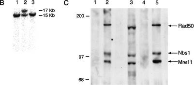
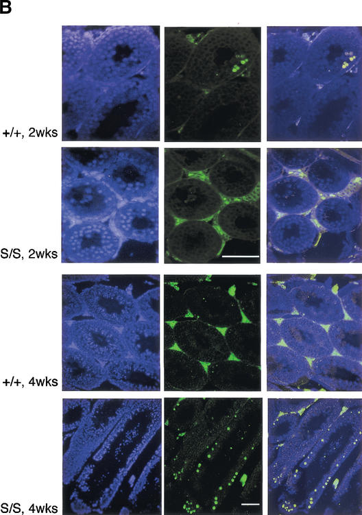
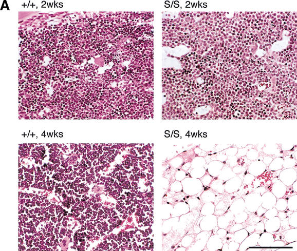
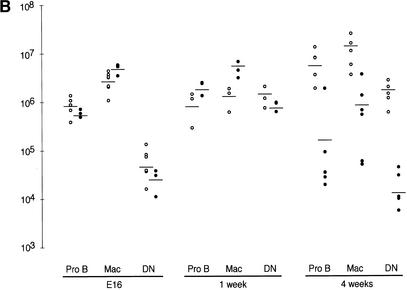
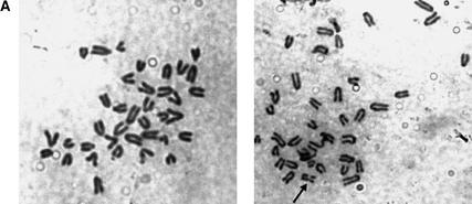
Mre11, Rad50, and Nbs1 function in a protein complex that is central to the metabolism of chromosome breaks. Null mutants of each are inviable. We demonstrate here that hypomorphic Rad50 mutant mice (Rad50(S/S) mice) exhibited growth defects and cancer predisposition. Rad50(S/S) mice died with complete bone marrow depletion as a result of progressive hematopoietic stem cell failure. Similar attrition occurred in spermatogenic cells. In both contexts, attrition was substantially mitigated by p53 deficiency, whereas the tumor latency of p53(-/-) and p53(+/-) animals was reduced by Rad50(S/S). Indices of genotoxic stress and chromosomal rearrangements were evident in Rad50(S/S) cultured cells, as well as in Rad50(S/S) and p53(-/-) Rad50(S/S) lymphomas, suggesting that the Rad50(S/S) phenotype was attributable to chromosomal instability. These outcomes were not associated with overt defects in the Mre11 complex's previously established double strand break repair and cell cycle checkpoint regulation functions. The data indicate that even subtle perturbation of Mre11 complex functions results in severe genotoxic stress, and that the complex is critically important for homeostasis of proliferative tissues.


Figures

Figure 1
Figure 1
Derivation of Rad50K22M mutants. (A) Schematic of Rad50 locus. 5′ region and exons 1–3 (black boxes) are shown. Homologous integration of the targeting vector pk-in-24ploxp with the K22M mutation encoded in exon 1 (*) inserts a loxP-flanked (triangles) puromyocin-resistance gene (Puro) antisense and 5′ of Rad50 and creates the Rad50K22M allele. Cre-mediated deletion of Puro creates the Rad50K22Mcre allele. N, NcoI; 5P, 5′ probe. (B) Southern blot analysis of NcoI-digested DNA hybridized with the 5′ probe shown in A. The Rad50 and Rad50K22M alleles yield 15 and 17 Kb fragments, respectively. (Lane 2) Targeted ES cell clone. (Lanes 1,3) Untargeted clones. (C) Rad50+/+ (lanes 1,2) or Rad50S/S (lanes 4,5) immunoprecipitations performed with preimmune (lanes 1,4) or Nbs1 antisera (lanes 2,5). Whole-cell extract of Rad50S/S MEFs (lane 3) included as control. Samples were immunoblotted sequentially with Nbs1, Mre11, and Rad50 antisera. Size markers (KD) are shown.
Figure 1
Figure 1
Derivation of Rad50K22M mutants. (A) Schematic of Rad50 locus. 5′ region and exons 1–3 (black boxes) are shown. Homologous integration of the targeting vector pk-in-24ploxp with the K22M mutation encoded in exon 1 (*) inserts a loxP-flanked (triangles) puromyocin-resistance gene (Puro) antisense and 5′ of Rad50 and creates the Rad50K22M allele. Cre-mediated deletion of Puro creates the Rad50K22Mcre allele. N, NcoI; 5P, 5′ probe. (B) Southern blot analysis of NcoI-digested DNA hybridized with the 5′ probe shown in A. The Rad50 and Rad50K22M alleles yield 15 and 17 Kb fragments, respectively. (Lane 2) Targeted ES cell clone. (Lanes 1,3) Untargeted clones. (C) Rad50+/+ (lanes 1,2) or Rad50S/S (lanes 4,5) immunoprecipitations performed with preimmune (lanes 1,4) or Nbs1 antisera (lanes 2,5). Whole-cell extract of Rad50S/S MEFs (lane 3) included as control. Samples were immunoblotted sequentially with Nbs1, Mre11, and Rad50 antisera. Size markers (KD) are shown.
Figure 2
Figure 2
Progressive degeneration in Rad50S/S testes is not associated with meiotic recombination defects. (A) Hematoxylin and eosin (H&E) staining of testes sections from 2- and 4-wk-old Rad50+/+ and Rad50S/S mice. Bar, 100 μm; magnification, 400×. (B) TUNEL-staining of apoptotic cells (FITC; green; middle) and nuclear counterstaining with DAPI (blue; left) in testes sections. Two-channel overlay of TUNEL-FITC and DAPI (right). Two-week-old sections, magnification 400×. Four-week-old sections, magnification 100×. Bars, 100 μm. (C) Surface-spread spermatocyte nuclei were stained with Cor1 antiserum, which recognizes Scp3 protein (red). Images of Rad50+/+ and Rad50S/S spermatocyte nuclei in pachytene demonstrate normal chromosome pairing. Chromosomes from a second meiotic cell are seen in the lower right corner of the Rad50S/S image. Bar, 10 μm; magnification, 1000×. Graph, meiotic progression in ≥200 Rad50+/+ (white bars) and Rad50S/S (black bars) nuclei determined by Cor1 staining patterns and chromosomal structure.
Figure 2
Figure 2
Progressive degeneration in Rad50S/S testes is not associated with meiotic recombination defects. (A) Hematoxylin and eosin (H&E) staining of testes sections from 2- and 4-wk-old Rad50+/+ and Rad50S/S mice. Bar, 100 μm; magnification, 400×. (B) TUNEL-staining of apoptotic cells (FITC; green; middle) and nuclear counterstaining with DAPI (blue; left) in testes sections. Two-channel overlay of TUNEL-FITC and DAPI (right). Two-week-old sections, magnification 400×. Four-week-old sections, magnification 100×. Bars, 100 μm. (C) Surface-spread spermatocyte nuclei were stained with Cor1 antiserum, which recognizes Scp3 protein (red). Images of Rad50+/+ and Rad50S/S spermatocyte nuclei in pachytene demonstrate normal chromosome pairing. Chromosomes from a second meiotic cell are seen in the lower right corner of the Rad50S/S image. Bar, 10 μm; magnification, 1000×. Graph, meiotic progression in ≥200 Rad50+/+ (white bars) and Rad50S/S (black bars) nuclei determined by Cor1 staining patterns and chromosomal structure.
Figure 3
Figure 3
Rad50S/S progressive hematopoietic depletion is intrinsic to stem cells. (A) H&E staining of bone marrow sections from Rad50+/+ and Rad50S/S. Bar, 100 μm; magnification, 400×. (B) FACS analysis of hematopoietic tissues from E16, 1-wk-old, and 4-wk-old mice. The number of Pro B cells, macrophages (Mac), and double negative T cells (DN) in Rad50+/+ (○) and Rad50S/S (●) mice is shown. Each symbol represents data for one mouse, and lines show the mean within each group. (C) FACS analysis of fetal liver cell transfer recipients 4–6 wk posttransfer. As in B; Rad50+/+ recipients of Rad50+/+ fetal liver cells (○), Rad50S/S recipients of Rad50+/+ cells (□), Rad50+/+ recipients of a 1:1 mixture of Rad50+/+ and Rad50S/S cells (▵), and Rad50+/+ recipients of Rad50S/S cells (●). Control Rad50+/+ mice that did not receive transfers died within 2 wk after irradiation.
Figure 3
Figure 3
Rad50S/S progressive hematopoietic depletion is intrinsic to stem cells. (A) H&E staining of bone marrow sections from Rad50+/+ and Rad50S/S. Bar, 100 μm; magnification, 400×. (B) FACS analysis of hematopoietic tissues from E16, 1-wk-old, and 4-wk-old mice. The number of Pro B cells, macrophages (Mac), and double negative T cells (DN) in Rad50+/+ (○) and Rad50S/S (●) mice is shown. Each symbol represents data for one mouse, and lines show the mean within each group. (C) FACS analysis of fetal liver cell transfer recipients 4–6 wk posttransfer. As in B; Rad50+/+ recipients of Rad50+/+ fetal liver cells (○), Rad50S/S recipients of Rad50+/+ cells (□), Rad50+/+ recipients of a 1:1 mixture of Rad50+/+ and Rad50S/S cells (▵), and Rad50+/+ recipients of Rad50S/S cells (●). Control Rad50+/+ mice that did not receive transfers died within 2 wk after irradiation.
Figure 4
Figure 4
Normal hairpin resolution in Rad50S/S thymocytes. Hairpin coding ends (CE), oligonucleotides were ligated to the D2/J1 blunt signal ends (SE) and the open CEs. Ends were PCR amplified using Dδ2– and oligospecific primers (arrows), and products were detected using the probe shown (line with a circle). Expected sizes of PCR products are indicated. Blot, LMPCR of 1 μg of MBN-treated thymic DNA from scid and Rad50S/S mice. M, radiolabeled size marker; sizes (bp) shown on left.
Figure 5
Figure 5
p53 deficiency partially rescues the Rad50S/S phenotype. (A) FACS analysis of 4-wk-old 129/SvEv Rad50+/+ (○), p53+/− Rad50S/S (▴), and p53−/−Rad50S/S (▪) mice; depicted as in Fig. 3B. (B) Mouse survival. Each data point represents the fraction of surviving mice at a given age in months. N, total number of mice for each genotype. Only autopsied mice are included. The average age of death in months (mo) is shown for each genotype with the P-value compared to the relevant control determined by the Wilcoxon rank sum test. (C) Results from TUNEL staining of testes sections from 4-mo-old mice. The % apoptotic tubules is the average fraction of tubules with ≥4 apoptotic cells in serial sections from two mice of each genotype.
Figure 5
Figure 5
p53 deficiency partially rescues the Rad50S/S phenotype. (A) FACS analysis of 4-wk-old 129/SvEv Rad50+/+ (○), p53+/− Rad50S/S (▴), and p53−/−Rad50S/S (▪) mice; depicted as in Fig. 3B. (B) Mouse survival. Each data point represents the fraction of surviving mice at a given age in months. N, total number of mice for each genotype. Only autopsied mice are included. The average age of death in months (mo) is shown for each genotype with the P-value compared to the relevant control determined by the Wilcoxon rank sum test. (C) Results from TUNEL staining of testes sections from 4-mo-old mice. The % apoptotic tubules is the average fraction of tubules with ≥4 apoptotic cells in serial sections from two mice of each genotype.
Figure 6
Figure 6
Chromosome instability in Rad50S/S cells. (A) Images of Giemsa-stained metaphase cells. (Left) a normal metaphase spread from a Rad50S/S ear fibroblast. (Right) Rad50S/S ear fibroblast metaphase with one broken chromosome (arrow). Magnification, 1000×. (B) SKY analysis of thymic lymphoma cells. Metaphase cells from p53−/−Rad50S/S (upper panel, tumor 5390) and Rad50S/S (lower panel, tumor 5587) tumors. (Left) Inverted image of the metaphase cells counterstained with DAPI. (Right) Spectral image of the metaphase cells. The structurally rearranged chromosomes are identified with arrows, and include short-arm fusions [der(13;14) and der(1;13)], an isodicentric fusion [idic(9)], and a small marker chromosome of unknown origin [mar] in the p53−/−Rad50S/S tumor, as well as dicentric and isodicentric rearrangements [dic(2;4) and idic(11)] in the Rad50S/S tumor. (C) (Right) Two-channel image of a representative metaphase spread from a p53−/−Rad50S/S lymphoma subjected to FISH using an FITC-conjugated telomeric probe (green) and counterstained with DAPI (blue). (Left) Inverted DAPI image of the metaphase cell. Bar, 10 μm; magnification, 1000×.
Figure 6
Figure 6
Chromosome instability in Rad50S/S cells. (A) Images of Giemsa-stained metaphase cells. (Left) a normal metaphase spread from a Rad50S/S ear fibroblast. (Right) Rad50S/S ear fibroblast metaphase with one broken chromosome (arrow). Magnification, 1000×. (B) SKY analysis of thymic lymphoma cells. Metaphase cells from p53−/−Rad50S/S (upper panel, tumor 5390) and Rad50S/S (lower panel, tumor 5587) tumors. (Left) Inverted image of the metaphase cells counterstained with DAPI. (Right) Spectral image of the metaphase cells. The structurally rearranged chromosomes are identified with arrows, and include short-arm fusions [der(13;14) and der(1;13)], an isodicentric fusion [idic(9)], and a small marker chromosome of unknown origin [mar] in the p53−/−Rad50S/S tumor, as well as dicentric and isodicentric rearrangements [dic(2;4) and idic(11)] in the Rad50S/S tumor. (C) (Right) Two-channel image of a representative metaphase spread from a p53−/−Rad50S/S lymphoma subjected to FISH using an FITC-conjugated telomeric probe (green) and counterstained with DAPI (blue). (Left) Inverted DAPI image of the metaphase cell. Bar, 10 μm; magnification, 1000×.

References

    1. Alani E, Padmore R, Kleckner N. Analysis of wild-type and rad50 mutants of yeast suggests an intimate relationship between meiotic chromosome synapsis and recombination. Cell. 1990;61:419–436. - PubMed
    1. Baudat F, Manova K, Yuen JP, Jasin M, Keeney S. Chromosome synapsis defects and sexually dimorphic meiotic progression in mice lacking Spo11. Mol Cell. 2000;6:989–998. - PubMed
    1. Bressan DA, Olivares HA, Nelms BE, Petrini JHJ. Alteration of N-terminal phosphoesterase signature motifs inactivates Saccharomyces cerevisiae Mre11. Genetics. 1998;150:592–600. - PMC - PubMed
    1. Bressan DA, Baxter BK, Petrini JHJ. The Mre11/Rad50/Xrs2 protein complex facilitates homologous recombination-based double strand break repair in Saccharomyces cerevisiae. Mol Cell Biol. 1999;19:7681–7687. - PMC - PubMed
    1. Carney JP, Maser RS, Olivares H, Davis EM, Le Beau M, Yates JR, 3rd, Hays L, Morgan WF, Petrini JHJ. The hMre11/hRad50 protein complex and Nijmegen breakage syndrome: Linkage of double-strand break repair to the cellular DNA damage response. Cell. 1998;93:477–486. - PubMed

Publication types

MeSH terms