. 2002 Sep;110(5):615-24.
doi: 10.1172/JCI15704.

Androgen receptor-mediated inhibition of cutaneous wound healing

Affiliations

Androgen receptor-mediated inhibition of cutaneous wound healing

Gillian S Ashcroft et al. J Clin Invest. 2002 Sep.

Abstract

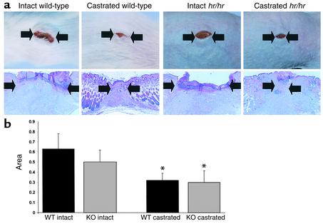
Impaired wound healing states in the elderly lead to substantial morbidity, mortality, and a cost to the US Health Services of over $9 billion per annum. In addition to intrinsic aging per se causing delayed healing, studies have suggested marked sex-differences in wound repair. We report that castration of male mice results in a striking acceleration of local cutaneous wound healing, and is associated with a reduced inflammatory response and increased hair growth. Using a hairless mouse model, we have demonstrated that testosterone reduction stimulates the healing response not through hair follicle epithelial/mesenchymal cell proliferation, but directly via effects on wound cell populations. We suggest that endogenous testosterone inhibits the cutaneous wound healing response in males and is associated with an enhanced inflammatory response. The mechanisms underlying the observed effects involve a direct upregulation of proinflammatory cytokine expression by macrophages in response to testosterone. Blockade of androgen action systemically, via receptor antagonism, accelerates healing significantly, suggesting a specific target for future therapeutic intervention in impaired wound healing states in elderly males.


Figures

Figure 1
Figure 1
AR localizes to keratinocytes, inflammatory cells, and fibroblasts during wound healing. (a) Normal skin immunostaining for AR (left panel) at low magnification (×20) illustrates epidermal and hair follicle staining. Right panels show high magnification (×100) of basal epidermal cells (area 1) and hair follicles (area 2). (b) Day 3 wounds: left panel is a low-magnification image of AR-stained tissue. Area 2 is a high-magnification image of macrophages, and area 1 shows basal and suprabasal epithelial staining at the migrating edge. (c) A day-14 wound (left panel, low magnification) with cells morphologically resembling fibroblasts staining positively for AR; (area 1, right panel) is a high-magnification image. Images are representative of ten wounds stained per timepoint from male mice. (d) AR-positive cells were quantified from day 1 to day 21 after wounding; those colocalizing with Mac-3 were quantified separately. (e) RT-PCR for AR and the housekeeping gene HPRT in wound tissue showed a temporal increase in expression through day 3, with a decrease by day 21. d0, normal, unwounded skin. Bottom panel shows a Western blot for AR (110 kDa) demonstrating an increase in protein levels from basal (d0) to day 7, then a decline in protein to day 21 after wounding. Blot is representative of three experiments using five mice per timepoint.
Figure 2
Figure 2
Accelerated healing in the absence of male gonadal steroids. (a) Left and central panels show macroscopic and histological day 5 wounds illustrating the smaller (healed) wounds in castrated animals compared with intact animals. Collagen content was markedly increased in the wounds of the castrated animals at day 5 (right panels). Magnification: ×10 (center, hematoxylin and eosin); ×20 (right, picrosirius red). (b) Cross-sectional wound areas were significantly reduced on days 3 and 7 after wounding in the castrated animals (n = 6–8 per group; *P < 0.05). (c) Hydroxyproline levels in wound tissue were significantly increased in the castrated mice compared with intact mice at days 5 and 21 after wounding. Extraction and analysis were performed on individual wounds from five mice per group at day 5, and on five samples of pooled wounds (three wounds per tube) at day 21 after wounding. Data represent mean value per 10 mg tissue. *P < 0.05.
Figure 3
Figure 3
Increased hair follicle proliferation following gonadectomy does not contribute to the wound healing response. (a) Day 3 macroscopic and histological wounds from intact wild-type and intact null (hr/hr) mice showed no differences in healing. By contrast, castrated hr/hr and castrated wild-type mice healed significantly more quickly. Magnification: ×10. (b) Quantification of wound areas at day 3 illustrated a significant decrease in area (accelerated healing) in the castrated hr/hr null (KO) animals compared with the intact hr/hr littermates, and wild-type (WT) castrated compared with wild-type intact. n = 6 per group; *P < 0.05.
Figure 4
Figure 4
Castration results in a dampened local inflammatory response and reduced expression of proinflammatory TNF-α. (a) Upper panels are low-magnification (×20) images of inflammatory cell staining for Mac-3. Mac-3–positive cells (arrow, bottom panels) were increased in day 3 wounds of intact mice compared with their castrated littermates (magnification: ×100). (b) Quantification of Mac-3–positive cells per unit area mm2 showed a significant increase in the intact animals at day 5 after wounding compared with castrated mice. (c) Expression of TNF-α was reduced at days 5 and 21 in the wounds of the castrated mice (C) compared with wounds of intact mice (I). Wound tissue was pooled from five mice, and the gel shown is representative of three experiments. Right panel illustrates a representative RNase protection assay (of three replicate experiments) showing no differences in tissue expression of IL-6, macrophage migration inhibitory factor (MIF), or TGF-β1 between intact and castrated mice. (d) TNF-α protein levels were increased in wounds of intact compared with castrated mice as illustrated by day 5 immunostaining (left panels, representative of six wounds stained per group). Quantification of immunostaining (graph) showed a significant increase at days 3, 5, and 21 in the wounds of the intact mice compared with the castrated mice. n = 6 per timepoint. Western blot analysis of wound tissue showed increased levels of TNF-α at days 5 and 21 in wounds of intact mice compared with those of castrated mice. Blot is representative of three experiments using five mice per group. *P < 0.05.
Figure 5
Figure 5
Sex differences in human wound healing. (a) Collagen VII immunostaining at day 14 after wounding illustrates complete reformation of the basement membrane in the young female (YF, left, arrow), whereas staining in the elderly female (EF) is patchy and punctate (green arrow), and is significantly fainter in the elderly male (EM, green arrow). NS, normal skin adjacent to wound. Young males healed similarly to young females, thus only the young female section is shown. (b) Quantification of collagen VII immunostaining where 0 = normal (unwounded) skin intensity. A significant (*P < 0.05) decrease in staining was apparent at day 14 in the elderly males. (c) TNF-α immunostaining at day 7 increased markedly with age, and dramatically in the elderly male. YM, young male. Arrows indicate positively staining macrophages. (d) Systemic testosterone levels in elderly health status–defined human males strongly correlates with impaired healing of acute wounds (increased wound area at day 7; P = 0.001; r2 = 0.459).
Figure 6
Figure 6
AR blockade accelerates healing and directly inhibits macrophage TNF-α expression. (a) Flutamide treatment significantly accelerates healing, as illustrated by reduced wound cross-sectional areas at day 3 after wounding (*P < 0.05) and by histological analysis (b) of day 3 wounds (arrows demarcate wound edge). (c) Reduced TNF-α expression at day 3 after wounding following flutamide treatment (F, oral flutamide; C, vehicle control; NS, normal skin from control animal). HPRT was used as the housekeeping gene. Bottom panel represents EMSA illustrating increased NF-κB binding activity in day 3 wounds (control) compared with normal skin; binding is markedly reduced in the day 3 wounds of flutamide-treated animals. (d) TGF-β1 (TGF)acted as a potent chemoattractant for murine peritoneal macrophages (C, control medium; *P < 0.05). Testosterone (Testo) had no effect on chemotaxis when used in conjunction with TGF-β1 or alone as a chemoattractant, nor when the cells were pretreated with testosterone. (e) LPS-induced TNF-α mRNA expression by murine macrophages. C, control; +, LPS. Testosterone markedly increased TNF-α expression both basally (T) and when cells were LPS-activated (T+). Flutamide treatment significantly reduced testosterone-induced TNF-α expression levels. Wound tissue was pooled from four mice, and the gel shown is representative of three experiments.

References

    1. Ashcroft GS, et al. Age-related differences in the temporal and spatial regulation of matrix metalloproteinases (MMPs) in normal skin and acute cutaneous wounds of healthy humans. Cell Tissue Res. 1997;290:581–591. - PubMed
    1. Herrick SE, et al. Up-regulation of elastase in acute wounds of healthy aged humans and chronic venous leg ulcers is associated with matrix degradation. Lab Invest. 1997;77:281–288. - PubMed
    1. Ashcroft GS, Horan MA, Ferguson MWJ. Aging alters the inflammatory and endothelial cell adhesion molecule profiles during human cutaneous wound healing. Lab Invest. 1998;78:47–58. - PubMed
    1. Ashcroft GS, Greenwell-Wild T, Horan MA, Wahl SM, Ferguson MWJ. Topical estrogen accelerates cutaneous wound healing in aged humans associated with an altered inflammatory response. Am J Pathol. 1999;155:1137–1146. - PMC - PubMed
    1. Taylor RJ, Taylor AD, Smyth JV. Using an artificial network to predict healing times and risk factors for venous leg ulcers. J Wound Care. 2002;11:101–105. - PubMed

Publication types

MeSH terms