. 2002 Sep;110(5):625-32.
doi: 10.1172/JCI15334.

Matrix metalloproteinases 2 and 9 work in concert to produce aortic aneurysms

Affiliations

Matrix metalloproteinases 2 and 9 work in concert to produce aortic aneurysms

G Matthew Longo et al. J Clin Invest. 2002 Sep.

Abstract

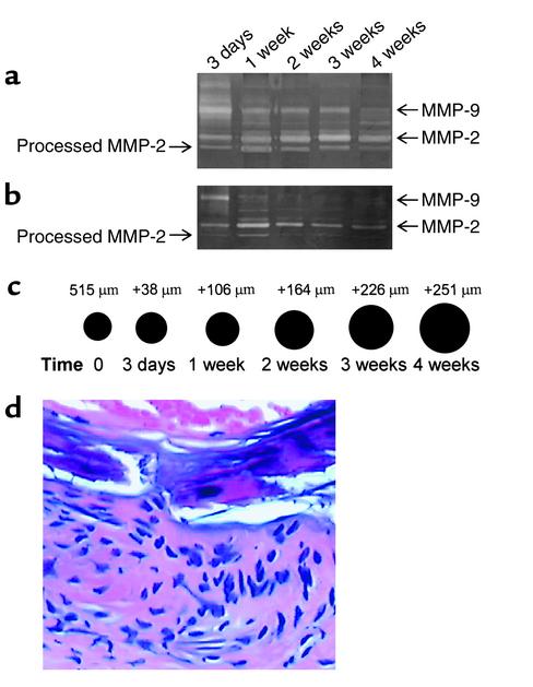
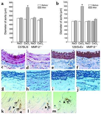
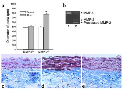
Matrix metalloproteinases (MMPs) 9 and 2 are increased in human abdominal aortic aneurysm (AAA) tissue, but their precise role and potential interaction remain unclear. Experimental induction of aortic aneurysms in mice genetically deficient in these peptidases could provide new insight into AAA pathogenesis. Mice deficient in the expression of MMP-9 (MMP-9KO) or MMP-2 (MMP-2KO) and their corresponding wild-type background mice (WT) underwent AAA induction by abluminal application of calcium chloride (CaCl(2)). No aneurysm formation was observed at 10 weeks after treatment in either the MMP-9KO or the MMP-2KO mice, whereas the corresponding WT mice showed an average 74% and 52% increase in aortic diameter, respectively. Reinfusion of competent macrophages from the corresponding WT strains into knockout mice resulted in reconstitution of AAA in MMP-9KO but not MMP-2KO mice. These findings suggest that macrophage-derived MMP-9 and mesenchymal cell MMP-2 are both required and work in concert to produce AAA.


Figures

Figure 1
Figure 1
The time course of expression of MMP-9 and MMP-2 and corresponding aneurysm growth. WT mouse aortae were incubated with CaCl2 or NaCl. Three days, 1 week, and 2, 3, and 4 weeks after treatment, the aortic diameters were measured and mice were sacrificed. (a) CaCl2-treated mice; (b) NaCl-treated mice (control). (c) Aortic diameter increases in CaCl2-treated mice at indicated time points. (d) H&E staining of CaCl2-treated mouse aortic section, showing a significant inflammatory infiltrate at 3 days.
Figure 2
Figure 2
(a and b) Aortic diameter changes after NaCl and CaCl2 treatment in C57BL/6 and MMP-2KO mice (a), and in 129/SvEv and MMP-9KO mice (b). Aortic diameters were measured before NaCl or CaCl2 incubation (white bars) and at sacrifice (gray bars). Bars shown in each group represent the mean ± SE of 5–14 mice. The diameter in the CaCl2-treated group was increased significantly at sacrifice in C57BL/6 and 129/SvEv mice (*P < 0.01). (cm) Histologic changes of mouse aorta at 10 weeks after treatment. (cf) Trichrome staining for elastic fibers. NaCl-treated (c) and CaCl2-treated (d) aorta from C57BL/6 and NaCl-treated (e) or CaCl2-treated (f) aorta from MMP-2KO mice. (gj) Pentachrome staining for elastic fibers. NaCl-treated (g) and CaCl2-treated (h) aorta from 129/SvEv and NaCl-treated (i) or CaCl2-treated (j) aorta from MMP-9KO mice. (km) Immunoperoxidase staining for macrophages of mouse aorta 2 weeks after CaCl2 incubation in 129/SvEv (k), MMP-9KO (l), and MMP-2KO (m). Mac3-positive cells are indicated by arrows. Elastic fibers are represented by arrowheads. (×100).
Figure 3
Figure 3
Gelatin zymography analysis of MMP activities expressed in experimental mice. Lane 1, WT; lanes 2 and 3, MMP-9KO; lanes 4 and 5, MMP-2KO.
Figure 4
Figure 4
(a) Aortic diameter changes after CaCl2 treatment with WT macrophage infusion in MMP-2KO and MMP-9KO mice. Aortic diameters were measured before (white bars) and 8 weeks after (gray bars) CaCl2 incubation. Bars shown in each group represent the mean ± SE of six to eight mice. The diameter in MMP-9KO mice increased significantly with macrophage infusion (*P < 0.01). (b) Gelatin zymography analysis of MMP activities expressed in WT peritoneal macrophages (lane 1) and aorta of MMP-9KO mouse infused with WT macrophages (lane 2). (ce) Histologic analysis. Paraffin sections were stained for elastin fibers (Trichrome), and sections were photographed and shown with the lumen at the top. (c) The MMP-9KO mouse infused with MMP-9KO macrophages; (d) the MMP-9KO mouse infused with WT macrophages; (e) the MMP-2KO mouse infused with WT macrophages.

Comment in

  • A confederacy of proteinases.
    Parks WC. Parks WC. J Clin Invest. 2002 Sep;110(5):613-4. doi: 10.1172/JCI16550. J Clin Invest. 2002. PMID: 12208861 Free PMC article. Review. No abstract available.

References

    1. Alcorn HG, Wolfson SK, Jr, Sutton-Tyrrell K, Kuller LH, O’Leary D. Risk factors for abdominal aortic aneurysms in older adults enrolled in The Cardiovascular Health Study. Arterioscler Thromb Vasc Biol. 1996;16:963–970. - PubMed
    1. Ernst CB. Abdominal aortic aneurysm. N Engl J Med. 1993;328:1167–1172. - PubMed
    1. Pearce WH, Koch AE. Cellular components and features of immune response in abdominal aortic aneurysms. Ann NY Acad Sci. 1996;800:175–185. - PubMed
    1. Bobryshev YV, Lord RS, Parsson H. Immunophenotypic analysis of the aortic aneurysm wall suggests that vascular dendritic cells are involved in immune responses. Cardiovasc Surg. 1998;6:240–249. - PubMed
    1. Anidjar S, et al. Elastase-induced experimental aneurysms in rats. Circulation. 1990;82:973–981. - PubMed

Publication types