. 2002 Sep;110(5):643-50.
doi: 10.1172/JCI16081.

An ATF2-derived peptide sensitizes melanomas to apoptosis and inhibits their growth and metastasis

Affiliations

An ATF2-derived peptide sensitizes melanomas to apoptosis and inhibits their growth and metastasis

Anindita Bhoumik et al. J Clin Invest. 2002 Sep.

Abstract

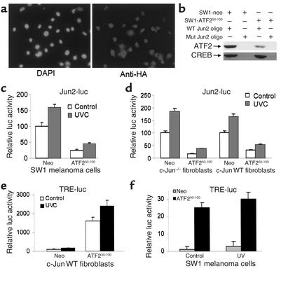
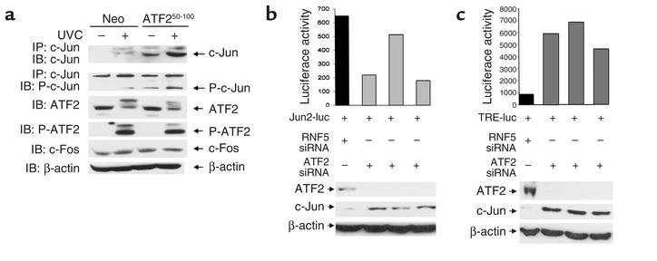
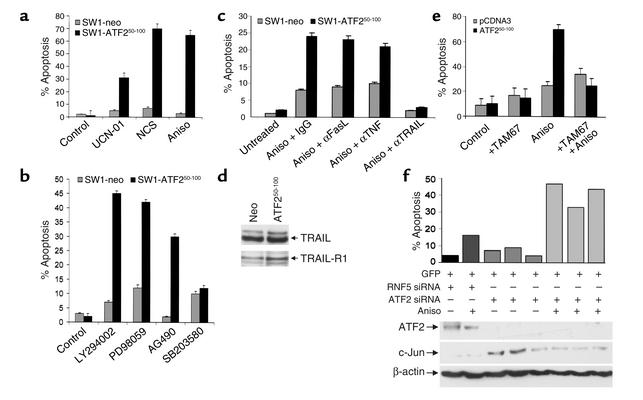
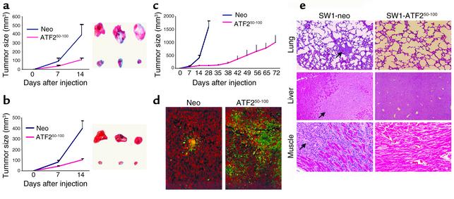
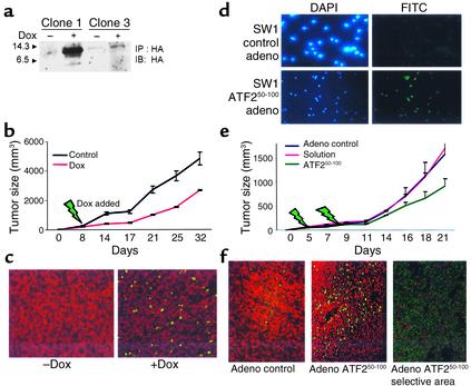
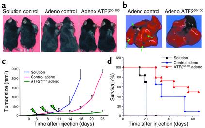
Melanomas are among the aggressive tumor types because of their notorious resistance to treatment and their high capacity to metastasize. ATF2 is among transcription factors implicated in the progression of melanoma and its resistance to treatment. Here we demonstrate that the expression of a peptide spanning amino acids 50-100 of ATF2 (ATF2(50-100)) reduces ATF2 transcriptional activities while increasing the expression and activity of c-Jun. Altering the balance of Jun/ATF2 transcriptional activities sensitized melanoma cells to apoptosis, an effect that could be attenuated by inhibiting c-Jun. Inhibition of ATF2 via RNA interference likewise increased c-Jun expression and primed melanoma cells to undergo apoptosis. Growth and metastasis of SW1 and B16F10 mouse melanomas were inhibited by ATF2(50-100) to varying degrees up to a complete regression, depending on the mode (inducible, constitutive, or adenoviral delivery) of its expression.


Figures

Figure 1
Figure 1
(a) Expression of the ATF250–100 peptide in SW1 cells. Mouse melanoma tumor-derived cells (SW1) were transfected with control or an HA-tagged ATF250–100 construct and clones were examined by immunohistochemistry using anti-HA antibodies. (b) Decreased ATF2 binding oligo bearing Jun2 motif in SW1 cells expressing ATF250–100 peptide. Nuclear proteins were incubated with biotinylated oligonucleotides bearing Jun2 motif. Oligo-bound proteins were captured on avidin-coated beads; then nonspecific bound proteins were removed by extensive washes, and bound material was subjected to analysis via Western blotting using antibodies to ATF2 and CREB. (c) Decreased Jun2-luc activity in SW1 mouse melanoma tumors expressing ATF250–100 peptide. SW1 cells that constitutively express control or ATF250–100 were transfected with the Jun2-luc and β-galactosidase (β-gal) constructs to monitor transcriptional activity of ATF2 and c-Jun. Proteins were assayed for β-gal activity and luciferase activity. Values depict relative luciferase activity normalized with respect to transfection efficiency based on β-gal assays. (d) Jun2-luc activity in Jun-null and WT cells. Shown is a Jun2-luc analysis similar to that shown in c except that the cell types used were Jun+/+ or null mouse fibroblasts. (e) TRE-luc activities in mouse fibroblast cells. Analysis of TRE-luc activities was carried out as indicated in c using mouse fibroblast cells that carry WT Jun. (f) SW1 cells expressing ATF2 peptide exhibit increased TRE-luc activity. Control and ATF2 peptide–expressing SW1 cells were transfected with the TRE-luc and β-gal constructs, and degree of TRE-mediated transcriptional activity was measured. Values were normalized per β-gal activity. DAPI, 4,6-diamino-2 phenylindole dilactate; UVC, ultraviolet-c radiation; neo, neomycin.
Figure 2
Figure 2
(a) Expression of ATF250–100 increases expression of c-Jun. Protein extracts prepared from indicated cells were subjected to immunoprecipitation with antibodies to c-Jun followed by immunoblot analysis using antibodies to phosphorylated c-Jun or control non-phosphoantibodies. Parallel analysis was carried out using antibodies to ATF2, c-Fos, and β-actin. Slow-migrating bands in ATF2 Western blots are likely to represent covalently modified forms of ATF2. (b) ATF2-siRNA alters c-Jun and ATF2 expression and the activity of Jun2-luc. SW1 cells were cotransfected with ATF2-siRNA and Jun2-luc as well as β-gal constructs. A portion of the same transfectants was taken for analysis of ATF2 and c-Jun expression to confirm inhibition of ATF2 by siRNA (lower panels). Luciferase assays carried out to monitor changes in TRE-mediated transcription are indicated following their normalization to β-gal. For the three experiments shown, P = 0.0167. (c) ATF2-siRNA alters c-Jun and ATF2 expression and the activity of TRE-luc. Experiment was performed as indicated in panel b, except that TRE-luc was used. For the three experiments shown, P = 0.0027. P-ATF2, phospho-ATF2; P-c-Jun, phospho-c-Jun.
Figure 3
Figure 3
(a) Sensitization of ATF250–100 peptide–expressing SW1 melanoma cells to apoptosis. SW1 cells expressing the ATF250–100 peptide or control peptide were treated with the indicated drugs, followed by FACS analysis to measure the hypodiploid cell populations. (b) Sensitization of SW1 melanoma to apoptosis by inhibitors of specific signaling pathways. SW1 cells expressing the ATF250–100 peptide were treated with the pharmacological inhibitors indicated, followed by FACS analysis to reveal the percentage of apoptotic cells. (c) Inhibition of TRAIL abolishes sensitivity of ATF250–100–expressing SW1 cells to anisomycin-induced apoptosis. Control and ATF250–100–expressing SW1 cells were pretreated with neutralizing antibodies to FasL, TRAIL, or TNF. Twenty-four hours later, cells were subjected to anisomycin (Aniso) treatment, and degree of apoptosis was monitored via FACS analysis. (d) ATF250–100–expressing cells exhibit an increase in expression of TRAIL receptor. Western blot analysis using antibodies to TRAIL or TRAIL receptor 1 was performed on extracts prepared from the indicated cells. (e) Dominant negative c-Jun construct attenuates the sensitivity of SW1 cells to anisomycin-induced apoptosis. Control cells and SW1 cells expressing ATF2 peptide were transfected with a dominant negative form of c-Jun (TAM67), and cells were subjected to anisomycin treatment. Degree of apoptosis was monitored via FACS analysis. (f) ATF2-siRNA sensitizes SW1 cells to apoptosis. Cotransfection of ATF2-siRNA and green fluorescent protein was followed by treatment of cells with anisomycin and analysis of green fluorescent protein–positive cells for apoptosis via FACS. For the three experiments shown, P = 0.0039. UCN-01, UCN-01-7-hydroxystaurosporine; NCS, neocarzinostatin.
Figure 4
Figure 4
(a) Mouse melanoma expressing the ATF250–100 peptide grow to a substantially smaller size. SW1 cells that express control vector or the ATF250–100 peptide were injected subcutaneously into C3H/HEJ mice, and tumor growth was monitored. The right panel depicts representative tumors at the end of the study, and the left panel shows tumor growth. The experiment was repeated three times. Bars = SD; P < 0.0001. (b) SW1 cells that express empty vector or the ATF250–100 peptide were injected subcutaneously into C3H/GLD (GLD mice bear mutations in FasL) mice, and follow-up and analysis were performed as indicated in a. Bars = SD; P < 0.0001. (c) Long-term growth of tumors from ATF250–100 peptide–expressing SW1 cells in C3H/HEJ mice. Tumors were measured at the indicated time points. Bars = SD; P < 0.0003. (d) Apoptosis of SW1 tumors expressing the ATF250–100 peptide. Sections of SW1 tumors produced in the presence of ATF250–100 peptide or control vector, tested for apoptosis by the TUNEL assay (apoptotic cells are green). (e) Expression of the ATF250–100 peptide in SW1 cells inhibits metastasis. Mice bearing tumors produced by cells that constitutively express the ATF250–100 peptide were monitored for metastatic lesions in the indicated organs at the end of inoculation. Arrows point to the metastatic lesions seen in the control group at the end of their inoculation.
Figure 5
Figure 5
Administration of ATF250–100 peptide to existing SW1 tumors reduces their growth in C3H mice. (ac) SW1 cells that exhibit inducible expression of the ATF250–100 peptide following addition of doxycycline were selected (a) and injected (clone 1) into C3H mice. Eight days after inoculation, doxycycline was added to the drinking water. Tumors were measured at the indicated times (b; P < 0.003). Analysis of apoptosis via the TUNEL assay revealed marked apoptosis in the tumors obtained from doxycycline-treated mice (c, green staining). (d) SW1 cells were infected with adenovirus (adeno) control or with adenovirus that expresses ATF250–100 peptide, and then subjected to immunohistochemistry analysis using antibodies to HA (HA is cloned in frame with the ATF2 peptide sequence). Left panels depict DAPI staining, whereas right panels show the fluorescence upon positive staining with HA antibodies. (e) SW1 cells were injected subcutaneously and tumors reached the size of 40 mm3 before virus control, solution control, or virus carrying the ATF2 peptide was injected (green arrows indicate injection). Data shown represent two experiments. P < 0.02 between control virus and the ATF2 peptide adenovirus groups. (f) Analysis of tumors for apoptosis via the TUNEL assay. Comparable magnification of control virus and ATF2 virus is shown in the two left panels; the right panel depicts a selective area representative of marked apoptosis within focal areas, seen in tumors that express the ATF2 peptide.
Figure 6
Figure 6
Intratumoral injection of adenovirus bearing the ATF2 peptide causes reduction of growth as well as complete regression of B16F10 tumors. C57BL/6 mice were injected with B16F10 cells, and tumors grew before injections of adenovirus bearing the ATF2 or control construct were initiated. Virus injection took place at the time points indicated in c (green arrows). (a) Representative tumors developed under each of the protocols. (b) The ATF2 peptide inhibited metastasis of the tumors. Green arrows point to metastatic lesions in the lungs seen in the control but not the treatment group; the black area in the photograph of ATF2 peptide–expressing mice depicts the heart. (c) Overall changes in the growth rate of the tumors subjected to control or ATF2 peptide treatments (compiled from the two experiments). Comparisons between all experimental groups are significant (P < 0.0001) as calculated by Tukey’s multiple comparisons (Tukey’s honestly significant difference). (d) The effect of ATF2 peptide on the survival of the mice under each of the protocols used.

References

    1. Yang YM, Dolan LR, Ronai Z. Expression of dominant negative CREB reduces resistance to radiation of human melanoma cells. Oncogene. 1996;12:2223–2233. - PubMed
    1. Jean D, Harbison M, McConkey DJ, Ronai Z, Bar-Eli M. CREB and its associated proteins act as survival factors for human melanoma cells. J. Biol Chem. 1998;273:24884–24890. - PubMed
    1. Yang J, Richmond A. Constitutive IkappaB kinase activity correlates with nuclear factor-kappaB activation in human melanoma cells. Cancer Res. 2001;61:4901–4909. - PubMed
    1. Recio JA, Merlino G. Hepatocyte growth factor/scatter factor activates proliferation in melanoma cells through p38 MAPK, ATF-2 and cyclin D1. Oncogene. 2002;21:1000–1008. - PubMed
    1. Ronai Z, et al. ATF2 confers radiation resistance to human melanoma cells. Oncogene. 1998;16:523–531. - PubMed

Publication types

MeSH terms