. 2002 Sep 2;196(5):589-604.
doi: 10.1084/jem.20020873.

Constitutive activation of the SRC family kinase Hck results in spontaneous pulmonary inflammation and an enhanced innate immune response

Affiliations

Constitutive activation of the SRC family kinase Hck results in spontaneous pulmonary inflammation and an enhanced innate immune response

Matthias Ernst et al. J Exp Med. .

Abstract

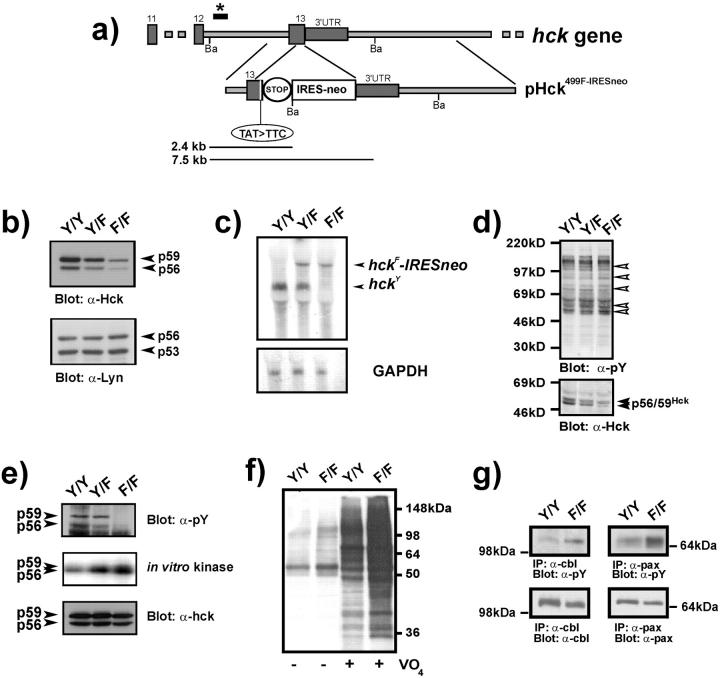
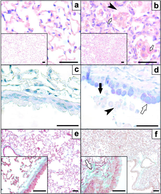
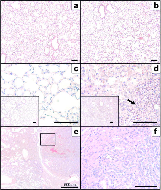
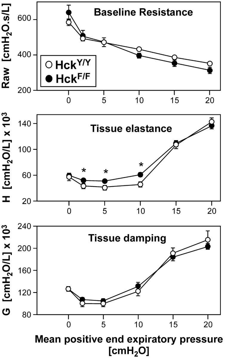
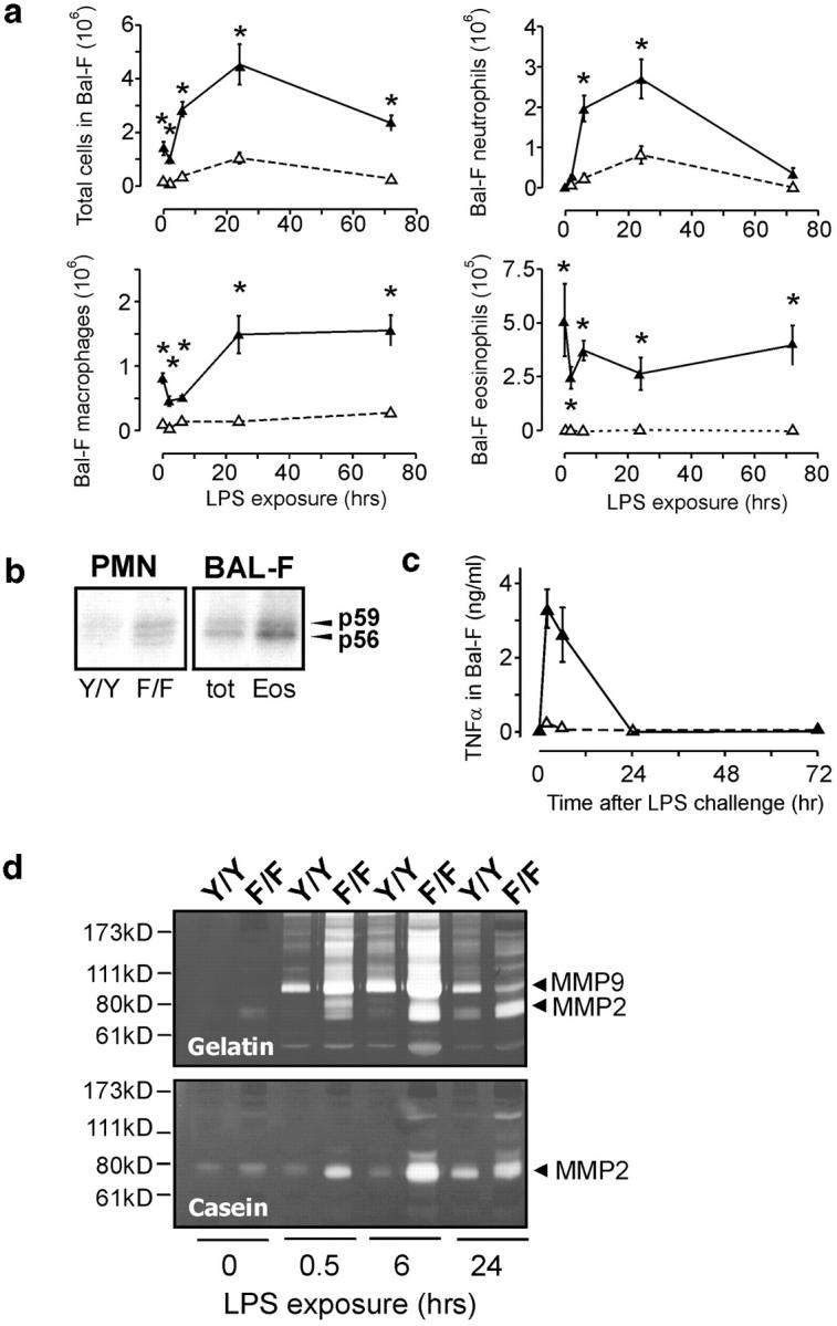
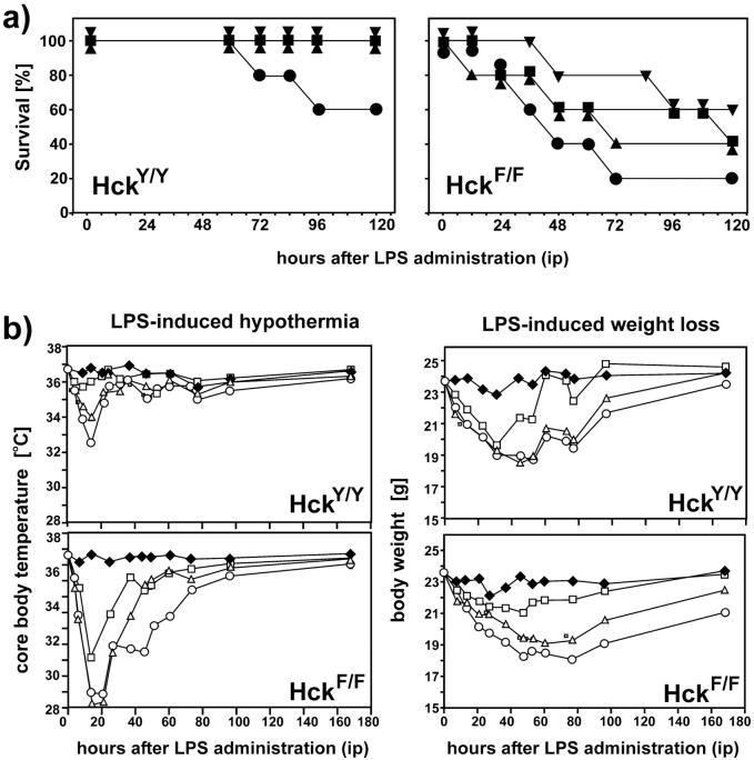
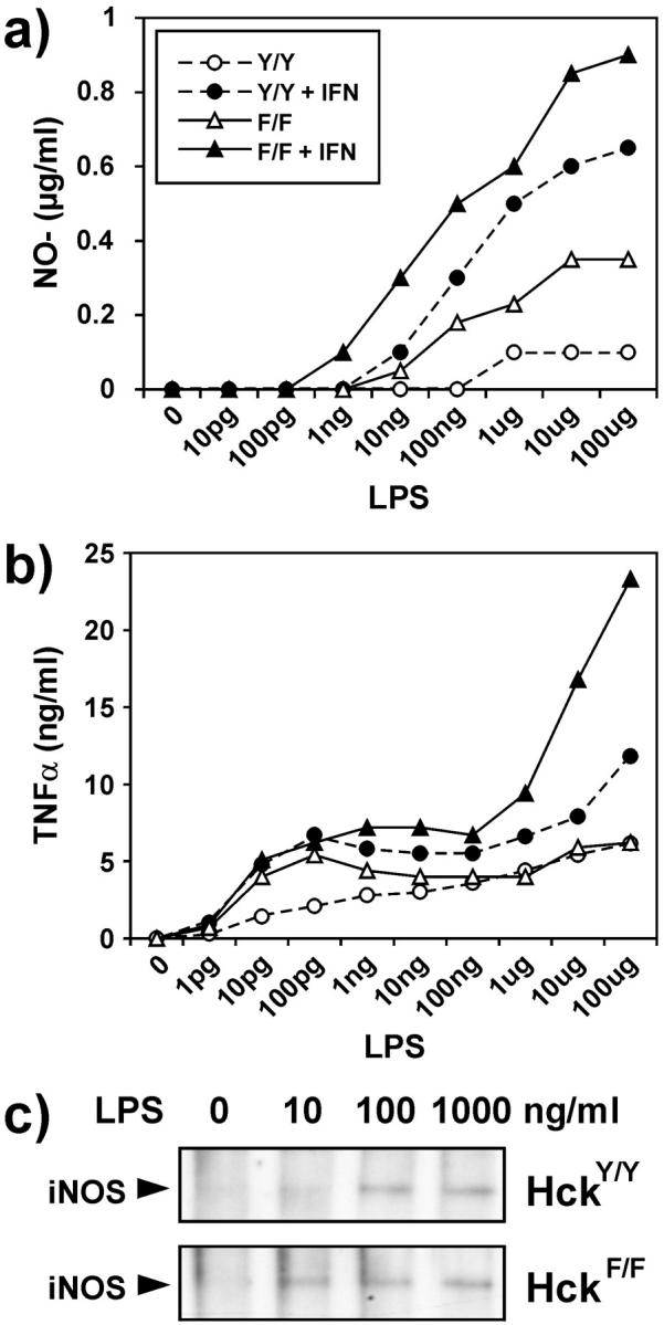
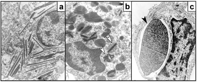
To identify the physiological role of Hck, a functionally redundant member of the Src family of tyrosine kinases expressed in myelomonocytic cells, we generated Hck(F/F) "knock-in" mice which carry a targeted tyrosine (Y) to phenylalanine (F) substitution of the COOH-terminal, negative regulatory Y(499)-residue in the Hck protein. Unlike their Hck(-/-) "loss-of-function" counterparts, Hck(F/F) "gain-of-function" mice spontaneously acquired a lung pathology characterized by extensive eosinophilic and mononuclear cell infiltration within the lung parenchyma, alveolar airspaces, and around blood vessels, as well as marked epithelial mucus metaplasia in conducting airways. Lungs from Hck(F/F) mice showed areas of mild emphysema and pulmonary fibrosis, which together with inflammation resulted in altered lung function and respiratory distress in aging mice. When challenged transnasally with lipopolysaccharide (LPS), Hck(F/F) mice displayed an exaggerated pulmonary innate immune response, characterized by excessive release of matrix metalloproteinases and tumor necrosis factor (TNF)alpha. Similarly, Hck(F/F) mice were highly sensitive to endotoxemia after systemic administration of LPS, and macrophages and neutrophils derived from Hck(F/F) mice exhibited enhanced effector functions in vitro (e.g., nitric oxide and TNFalpha production, chemotaxis, and degranulation). Based on the demonstrated functional association of Hck with leukocyte integrins, we propose that constitutive activation of Hck may mimic adhesion-dependent priming of leukocytes. Thus, our observations collectively suggest an enhanced innate immune response in Hck(F/F) mice thereby skewing innate immunity from a reversible physiological host defense response to one causing irreversible tissue damage.


Figures

Figure 1.
Figure 1.
Generation of HckF/F mice and biochemical analysis of HckF/F BMDMs. (a) Gene targeting strategy for introducing the Y499F mutation into the endogenous hck gene in ES cells. A region of the murine hck gene is schematically depicted with numbers of the coding exons (boxes) and the 3-UTR. The targeting vector pHCK499F-IRESneo contains the TAT to TTC substitution in the nucleotide triplet encoding amino acid 499, a stop codon, and an IRES-neomycin resistance (IRESneo) cassette. A diagnostic digest with BamHI (Ba) yields a fragment of 7.5 kb from the wild-type allele and a 2.4-kb fragment from the targeted allele when hybridized with a probe external to the targeting vector (marked by *). (b) Reduced levels of HckF protein in BMDM. Equal amounts of total cell lysates derived from mice of the indicated genotypes were subjected to Western blotting with anti-Hck antibody. The blot was stripped and re-probed with an anti-Lyn antibody. Arrows indicate the positions of the p59/p56 Hck and p56/p53 Lyn isoforms, respectively. (c) Northern blot analysis of BMDM RNA. Poly(A)+RNA was hybridized with a full-length cDNA clone encoding murine Hck. Dicistronic hck F-IRESneo mRNA transcripts are ∼1.4 kb larger than the wild-type hck mRNA transcripts. A gapdh probe was subsequently used to assess RNA loading. (d) Elevated levels of tyrosine-phosphorylated proteins in HckF/F mutant cells. Total cell lysates from BMDMs derived from mice of the indicated genotypes were subjected to Western blotting with anti-phosphotyrosine (pY) antibodies. The blot was then stripped and reprobed with an anti-Hck antibody. (e) HckF exhibits elevated activity in vitro. Equalized amounts of Hck were immunoprecipitated from lysates of BMDMs (bottom panel) and subjected to either Western blotting with anti-phosphotyrosine (pY) antibody (top panel) or an auto-phosphorylation assay in the presence of γ-[32P]-ATP (middle panel). (f) Enhanced tyrosine-phosphorylation in BMDMs in the presence of pervanadate. Total cell lysates prepared from BMDMs that had been cultured in the presence or absence of 1.5 μM pervanadate (VO4) were subjected to Western blotting with an anti-pY antibody. (g) The lysates used in f were also used to analyze tyrosine-phosphorylation of the Hck substrates c-Cbl and paxillin. 1 mg of total cell lysate was immunoprecipitated with the indicated antibody and then subjected to Western blotting with an anti-pY antibody. Stripped blots were subsequently reprobed with anti-Cbl and anti-paxillin antibodies to confirm equal protein loading.
Figure 2.
Figure 2.
Histological analysis of lungs from HckF/F mice. Cross sections of lungs collected from HckY/Y mice (a, c, and e) and HckF/F mice (b, d, and f) at 3 mo of age. Mutant lungs consistently showed accumulation of monocytes and macrophages in the alveolar airspaces in lungs of HckF/F mice (b and f). Alveolar macrophages in mutant lungs were frequently vacuolated (b and d; arrowheads) with eosinophilic cytoplasm (panel b inset; arrows). Epithelial cells lining the conducting airways of mutant mice displayed extensive mucus cell metaplasia (blue/purple stain in d; white arrow) resulting in mucus aggregates in the airway lumen (d, black arrow) when compared with wild-type mice (c). Mutant lungs were characterized by excessive fibrotic deposits of extracellular matrix in the alveolar septa (pale green stain in f, arrow). Histological stains were hematoxylin and eosin (a and b), alcian blue/periodic acid-Schiff (c and d), or Masson's trichrome (e and f). Bar = 40 μm.
Figure 3.
Figure 3.
Acquired pulmonary changes in HckF/F mice. Cross sections of lungs collected from HckY/Y mice (a and c) and HckF/F mice (b, d, e, and f). The lungs of mutant mice look histologically indistinguishable at weaning age (a and b), but show the characteristic accumulation of macrophages and eosinophils (arrow) by 6 wk of age (c and d). Occasionally, old mutant mice (∼2 yr of age) develop pulmonary adenocarcinoma (e and f). Panel f represents a magnification of the boxed area in e and shows well-differentiated adenocarcinoma composed of irregular glands lined by neoplastic epithelial cells (arrowhead). Note the extensive consolidation in the nonmalignant sections of the lung (e). Histological stains were hematoxylin and eosin. Bar = 100 μm.
Figure 4.
Figure 4.
Impaired lung function in HckF/F mice. Lung function analysis by oscillatory mechanics indicated that despite intense inflammation and incipient emphysema, HckF/F mice did not have increased baseline resistance (Raw) or marked altered tissue damping (G) when compared with wild-type mice. However, mutant lungs were stiffer (increased tissue elastance, H), particularly at low lung volumes, possibly resulting from extensive pulmonary consolidation. Mean ± SD (n = 6); *P < 0.05.
Figure 5.
Figure 5.
Enhanced pulmonary innate immune response in HckF/F mice. (a) Cellular composition of BAL-F cells from mutant HckF/F (filled symbols) and wild-type HckY/Y mice (open symbols) of 10 wk of age after transnasal challenge with LPS (10 μg/mouse). BAL-F was collected as described in Materials and Methods and cells from eight mice were counted and classified by their morphological appearance in cytospins. Mean ± SD, *P < 0.01. Data were analyzed by oneway analysis of variance (ANOVA) followed by Newman-Kreuls multiple comparison tests. (b) In vitro Hck-kinase activity of leukocytes. PMNs were collected from the bone-marrow of adult HckF/F and HckY/Y mice, whereas total BAL-F cells came only from lungs of unchallenged adult HckF/F mice, as BAL-F of HckY/Y contains negligible numbers of eosinophils. The eosinophil-enriched (Eos) population represented the lectin-bound proportion of total BAL-F cells (tot) as described in Materials and Methods. 500 μg of cell lysate was then immunoprecipitated with an anti-Hck antibody and in vitro autokinase assays were performed in the presence of γ-[32P]-ATP. (c) TNFα levels in BAL-F from LPS-challenged mice. After a single transnasal challenge with LPS (10 μg/mouse), levels of TNFα in BAL-F from HckF/F and HckY/Y mice (three mice of each genotype per experiment) were measured. Filled symbols: HckF/F mice, open symbols: HckY/Y mice. (d) MMP-9 and MMP-2 activity in BAL-F from LPS-challenged mice. After a single transnasal challenge with LPS (10 μg/mouse), BAL-F was recovered from the mice at the indicated time points and MMP-9 and MMP-2 activity was assayed by zymography using gelatin and casein as the respective substrates. Zones of MMP activity appear as unstained bands corresponding to the apparent molecular weights of MMP-2 and MMP-9, respectively.
Figure 6.
Figure 6.
Enhanced susceptibility of HckF/F mice to systemic LPS-challenges. (a) Reduced survival of HckF/F mutant mice compared with HckY/Y wild-type mice after a single intraperitoneal injection of LPS at 6 mg/kg (▾); 12 mg/kg (▪); 16 mg/kg (▴) or 24 mg/kg (•) and a 5 d observation period for survival. Five mice were used per treatment group. (b) Enhanced morbidity of HckF/F mice in response to administration of sublethal amounts of LPS. 3 d before LPS administration, a telemetric temperature-measuring device was implanted subcutaneously in the back of mice and temperature and weight recorded at the indicated time points after a single intraperitoneal injection of LPS at 2 mg/kg (U); 4 mg/kg (Δ); 8 mg/kg (○) or vehicle (♦). Each curve represents the median of five mice per group, except for the 8 mg/kg HckF/F group which only depicts the results from the three surviving mice.
Figure 7.
Figure 7.
Exaggerated in vitro response of BMDMs to LPS. (a and b) Release of NO2 (a) and TNFα (b) by BMDMs, derived from 8-wk-old HckF/F and HckY/Y mice and exposed to the indicated amount of LPS for 48 h. Where indicated, the cultures were primed with IFN-γ (15 U/ml) 2 h before LPS treatment. Values are the median of quadruplicate cultures. (c) Induction of nitric oxide synthetase protein (iNOS) in LPS-treated BMDMs of HckF/F and HckY/Y mice was assessed by Western blotting of 50 μg of total cell lysates after a 24-h stimulation with LPS at the indicated concentration.
Figure 8.
Figure 8.
Enhanced phagocytic activity of HckF/F macrophages. (a–c) Electron micrographs of alveolar macrophages from HckF/F mice showing the presence of eosinophilic crystals (a, original magnification ×25,000), which are often associated with secondary lysosomes (b; arrowhead, original magnification ×50,500), and frequently contain ingested erythrocytes (c; arrowhead, original magnification ×25,000). (d) Half-life of erythrocytes in HckF/F and HckY/Y mice. Levels of 51Cr-labeled erythrocytes in the peripheral blood of HckF/F and HckY/Y mice were determined at the indicated time points after transfusion. The amount of radioactivity associated with erythrocyte 30 min after transfusion was taken as 100%. The data represent means ± SD (n = 10). (e) Phagocytosis of SRBCs by BMDMs. Nonopsonized or anti-SRBC antiserum opsonized 51Cr-labeled SRBC were added to cultures of BMDMs derived from HckF/F or HckY/Y mice. After a 2-h incubation, the cultures were washed extensively and radioactivity released after lysis of the BMDMs was expressed as a proportion of total radioactivity added to each well. Data represent mean ± SD of quadruplicate cultures from a representative experiment.
Figure 8.
Figure 8.
Enhanced phagocytic activity of HckF/F macrophages. (a–c) Electron micrographs of alveolar macrophages from HckF/F mice showing the presence of eosinophilic crystals (a, original magnification ×25,000), which are often associated with secondary lysosomes (b; arrowhead, original magnification ×50,500), and frequently contain ingested erythrocytes (c; arrowhead, original magnification ×25,000). (d) Half-life of erythrocytes in HckF/F and HckY/Y mice. Levels of 51Cr-labeled erythrocytes in the peripheral blood of HckF/F and HckY/Y mice were determined at the indicated time points after transfusion. The amount of radioactivity associated with erythrocyte 30 min after transfusion was taken as 100%. The data represent means ± SD (n = 10). (e) Phagocytosis of SRBCs by BMDMs. Nonopsonized or anti-SRBC antiserum opsonized 51Cr-labeled SRBC were added to cultures of BMDMs derived from HckF/F or HckY/Y mice. After a 2-h incubation, the cultures were washed extensively and radioactivity released after lysis of the BMDMs was expressed as a proportion of total radioactivity added to each well. Data represent mean ± SD of quadruplicate cultures from a representative experiment.
Figure 9.
Figure 9.
Enhanced adhesion-dependent activity of HckF/F PMNs in vitro. (a) PMNs from HckF/F (black bars) and HckY/Y (white bars) mice were seeded in the top well of a modified Boyden chamber containing a fibronectin-coated insert studded with 8-μM pores. The chemoattractant fMLP (200 nM) as added to lower and/or upper well as indicated. After incubation at 37°C for the indicated periods of time, the inserts were removed, stained with Giemsa and the number of PMNs that had migrated to the underside of the inserts (left panels) were quantified by counting the number of cells in five separate fields on two inserts per time point (right panel). Mean ± SD. (b) Activation and degranulation response of Gr-1–positive PMNs as determined by FACS® analysis. HckY/Y (white symbols) and HckF/F PMNs (black symbols) were either preincubated for 30 min at 37°C in fibrinogen-coated 24-well plates (“adherent”) or directly stimulated (“suspension”) with TNFα (20 ng/ml) or PMA (1 mg/ml). Cells were fixed at the indicated time points, stained for Gr-1, and changes in forward scatter (FSC) and side scatter (SSC) profiles was analyzed (left panels). The percentage of Gr-1hi cells with reduced SSC (“degranulation”) and increased FSC (“activation”) were gated in the bottom right quadrant. The data shown are shown from a representative experiment (right panel). (c) Generation of O2 is exaggerated in HckF/F PMNs. PMNs from HckY/Y (white symbols) and HckF/F (black symbols) mice were seeded into fibrinogen-coated 96-well plates. The cells were stimulated with TNFα (20 ng/ml), fMLP (200 nM), or vehicle and incubated at 37°C for the indicated periods of time. O2 production was then measured as described in Materials and Methods. The data represents means ± SD from quadruplicate cultures.

References

    1. Kefalas, P., T.R. Brown, and P.M. Bricknell. 1995. Signalling by the p60c-src family of protein-tyrosine kinases. Int. J. Biochem. Cell Biol. 27:551–563. - PubMed
    1. Sicheri, F., and J. Kuriyan. 1997. Structures of Src-family tyrosine kinases. Curr. Opin. Struct. Biol. 7:777–785. - PubMed
    1. Cartwright, C.A., W. Eckhart, S. Simon, and P.L. Kaplan. 1987. Cell transformation by pp60c-src mutated in the carboxy-terminal regulatory domain. Cell. 49:83–91. - PubMed
    1. Kmiecik, T.T., and D. Shalloway. 1987. Activation and suppression of pp60c-src transforming ability by mutation of its primary sites of tyrosine phosphorylation. Cell. 49:65–73. - PubMed
    1. Piwnica-Worms, H., K.B. Saunders, T.M. Roberts, A.E. Smith, and S.H. Cheng. 1987. Tyrosine phosphorylation regulates the biochemical and biological properties of pp60c-src. Cell. 49:75–82. - PubMed

MeSH terms