. 2002 Oct;76(19):9868-76.
doi: 10.1128/jvi.76.19.9868-9876.2002.

Memory CD4(+) T cells are the earliest detectable human immunodeficiency virus type 1 (HIV-1)-infected cells in the female genital mucosal tissue during HIV-1 transmission in an organ culture system

Affiliations

Memory CD4(+) T cells are the earliest detectable human immunodeficiency virus type 1 (HIV-1)-infected cells in the female genital mucosal tissue during HIV-1 transmission in an organ culture system

Phalguni Gupta et al. J Virol. 2002 Oct.

Abstract

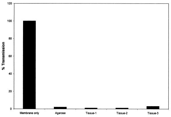
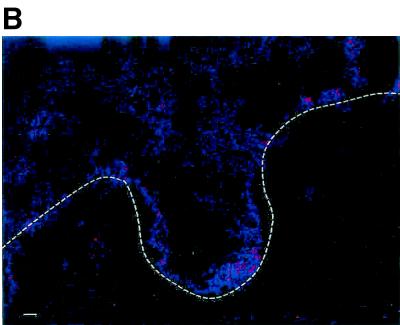
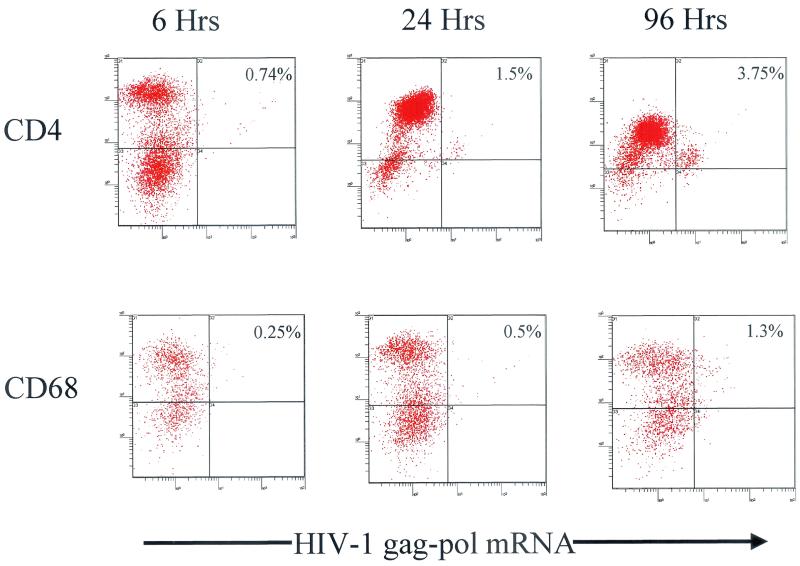
The virologic and cellular factors that are involved in transmission of human immunodeficiency virus type 1 (HIV-1) across the female genital tissue are poorly understood. We have recently developed a human cervical tissue-derived organ culture model to study heterosexual transmission of HIV-1 that mimics the in vivo situation. Using this model we investigated the role of phenotypic characteristics of HIV-1 and identified the cell types that are first infected during transmission. Our data indicate that the cell-free R5 HIV-1 was more efficiently transmitted than cell-free X4 HIV-1. Cell-free and cell-associated HIV-1 had comparable transmission efficiency regardless of whether the virus was of R5 or X4 type. We have demonstrated that memory CD4(+) T cells and not Langerhans cells were the first HIV-1 RNA-positive cells detected at the epithelial-submucosal junction 6 h after virus exposure. Multicolor laser confocal microscopy demonstrated a globular distribution of HIV-1 gag-pol mRNA in the cytoplasm, and the distribution of CD4 and the CD45RO isoform was irregular on the cellular membrane. At 96 h postinoculation, in addition to memory CD4(+) T cells, HIV-1 RNA-positive Langerhans cells and macrophages were also detected. The identification of CD4(+) T cells in the tissue at 6 h was confirmed by flow cytometric simultaneous immunophenotyping and ultrasensitive fluorescence in situ hybridization assay on immune cells isolated from disaggregated tissue. Furthermore, PMPA [9-[2-(phosphonomethoxy)propyl] adenine], an antiretroviral compound, and UC781, a microbicide, inhibited HIV-1 transmission across the mucosa, indicating the utility of the organ culture to screen topical microbicides for their ability to block sexual transmission of HIV-1.


Figures

FIG. 1.
FIG. 1.
Transmission of fluorescently labeled beads across the cervical tissue after 24 h in culture. Transmission was measured by flow cytometry of the culture medium from the bottom well.
FIG. 2.
FIG. 2.
Transmission electron micrograph of cervical tissue at day 0 and after 1 and 3 days of culture in our standard organ culture condition. The arrow indicates the basal layer junction between the epithelial layer and the squamous cell layer.
FIG. 3.
FIG. 3.
Kinetics of HIV-1 transmission from cell-free and cell-associated virus across the mucosa of the cervical tissue. (A) Cell-free BAL; (B) PBMC infected with BAL; (C) cell-free IIIB; (D) PBMC infected with IIIB. Percentage of transmission was calculated by the number of blue-stained cMAGI cells obtained from the culture supernatant of the tissue well divided by the number of blue-stained cMAGI cells obtained from the supernatant of the membrane-only well.
FIG. 4.
FIG. 4.
Identification of cell types that first become infected in cervical tissue during transmission of HIV-1. Tissues from a single patient were exposed to cell-free HIV-1 BAL for 6, 24, and 96 h and then analyzed for the identification of cell types expressing HIV-1 gag-pol mRNA by simultaneous two-parameter immunophenotyping and ultrasensitive fluorescence in situ hybridization as previously described (3, 22). (A) Tissues stained with CD4, CD45, and CD1a antibodies and hybridized with HIV-1 gag-pol probes. T cells are indicated by arrows, and Langerhans cells are indicated by arrowheads. The inset (scale bar = 10 μm) is a three-dimensional, three-parameter, laser confocal image of a productively infected CD4+ CD45RO+ T cell at 24 h postinfection. This infected cell demonstrated a globular distribution of HIV-1 gag-pol mRNA (green) in the cytoplasm. Further, the distribution of CD4 (red) and the CD45RO (blue) isoform was irregular on the cell membrane. (B) Control tissue from the same individual as in panel A, stained with anti-immunoglobulin-phycoerythrin and anti-immunoglobulin-Cy5, and hybridized under the same conditions as in panel A with a sense probe cocktail. Scale bar, 20 μm. The dashed line indicates the epithelial-submucosal junction.
FIG. 4.
FIG. 4.
Identification of cell types that first become infected in cervical tissue during transmission of HIV-1. Tissues from a single patient were exposed to cell-free HIV-1 BAL for 6, 24, and 96 h and then analyzed for the identification of cell types expressing HIV-1 gag-pol mRNA by simultaneous two-parameter immunophenotyping and ultrasensitive fluorescence in situ hybridization as previously described (3, 22). (A) Tissues stained with CD4, CD45, and CD1a antibodies and hybridized with HIV-1 gag-pol probes. T cells are indicated by arrows, and Langerhans cells are indicated by arrowheads. The inset (scale bar = 10 μm) is a three-dimensional, three-parameter, laser confocal image of a productively infected CD4+ CD45RO+ T cell at 24 h postinfection. This infected cell demonstrated a globular distribution of HIV-1 gag-pol mRNA (green) in the cytoplasm. Further, the distribution of CD4 (red) and the CD45RO (blue) isoform was irregular on the cell membrane. (B) Control tissue from the same individual as in panel A, stained with anti-immunoglobulin-phycoerythrin and anti-immunoglobulin-Cy5, and hybridized under the same conditions as in panel A with a sense probe cocktail. Scale bar, 20 μm. The dashed line indicates the epithelial-submucosal junction.
FIG. 5.
FIG. 5.
Representative two-color dot plots of productively infected T cells and macrophages from cells isolated from cervical explant tissue postinfection at 6, 24, and 96 h. The background signal was 0.3%.
FIG. 6.
FIG. 6.
Effect of antiretroviral compound PMPA (A) and microbicide UC781 (B) on the transmission of cell-free HIV-1 BAL across the genital mucosa. The data represent the number of blue-stained cells at the peak point (day 1).

References

    1. Balachandran, R., P. Thampatty, A. Enrico, C. Rinaldo, and P. Gupta. 1991. Human immunideficiency virus isolates from asymptomatic homosexual men and from AIDS patients have distinct biologic and genetic properties. Virology 180:229-238. - PubMed
    1. Balzarini, J., L. Naesens, E. Verbeken, M. Laga, L. Van Damme, M. Parniak, L. Van Mellaert, J. Anne, and E. De Clercq. 1998. Preclinical studies on thiocarboxanilide UC781 as a virucidal agent. AIDS 12:1129-1138. - PubMed
    1. Behbahani, H., E. Popck, P. Garcia, J. Anderson, A. L. Spectz, A. Landay, Z. Flener, and B. K. Patterson. 2000. Upregulation of CCR5 expression in the placenta is associated with human immunodeficiency virus-1 vertical transmission. Am. J. Pathol. 157:1811-1818. - PMC - PubMed
    1. Borkow, G., J. Barnard, T. M. Nguyen, A. Belmonte, M. A. Wainberg, and M. A. Parniak. 1997. Chemical barrier to human immunodeficiency virus type 1 (HIV-1) infection: retrovirucidal activity of UC781, a thiocarboxanilide nonnucleoside inhibitor of HIV-1 reverse transcriptase. J. Virol. 71:3023-3030. - PMC - PubMed
    1. Chackerian, B., E. Long, P. Luciw, and J. Overbaugh. 1997. Human immunodeficiency virus type 1 coreceptors participate in postentry stages of the virus replication cycle and function in simian immunodeficiency virus infection. J. Virol. 71:3932-3939. - PMC - PubMed

Publication types

Substances