. 2002 Oct;76(19):9920-33.
doi: 10.1128/jvi.76.19.9920-9933.2002.

Strategy for nonenveloped virus entry: a hydrophobic conformer of the reovirus membrane penetration protein micro 1 mediates membrane disruption

Affiliations

Strategy for nonenveloped virus entry: a hydrophobic conformer of the reovirus membrane penetration protein micro 1 mediates membrane disruption

Kartik Chandran et al. J Virol. 2002 Oct.

Abstract

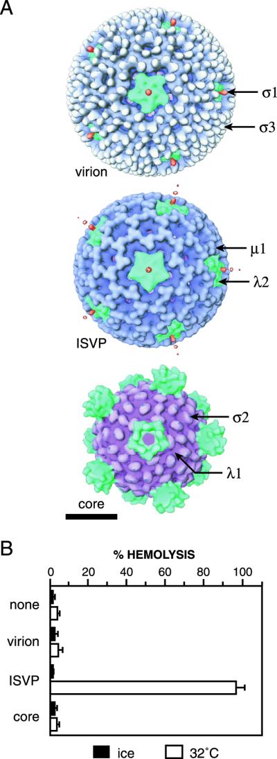
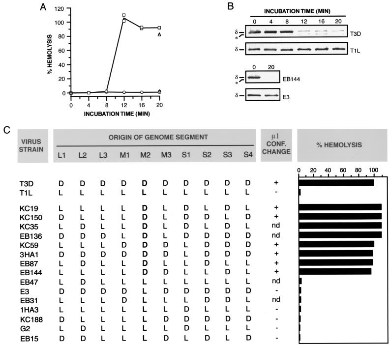
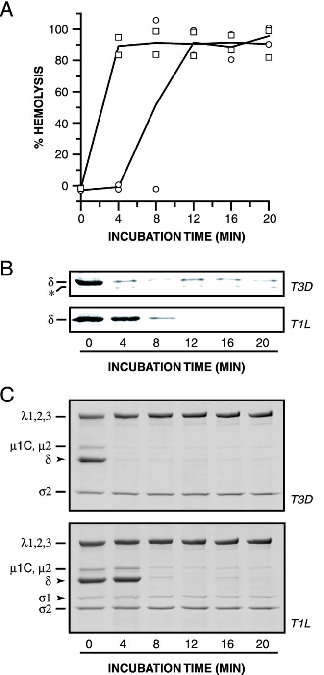
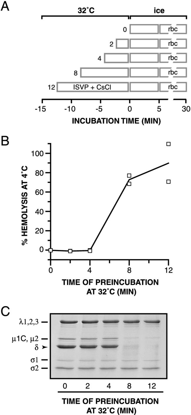
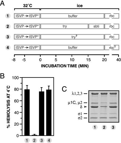
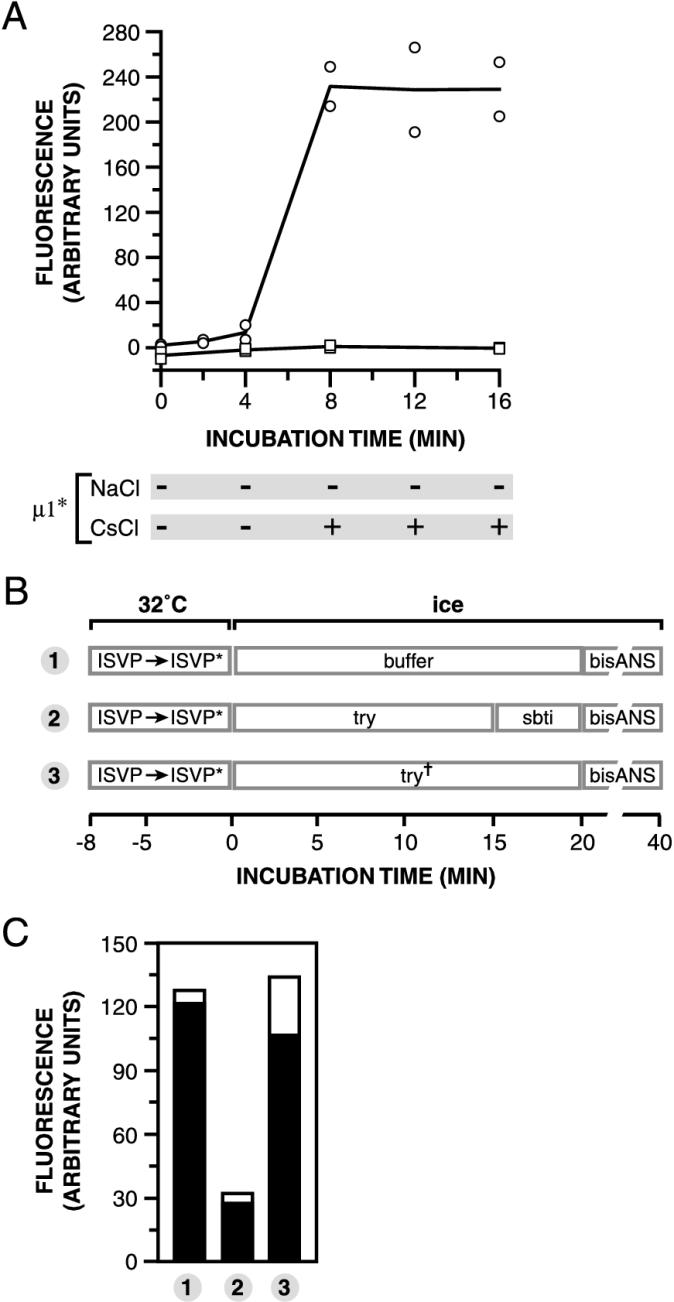
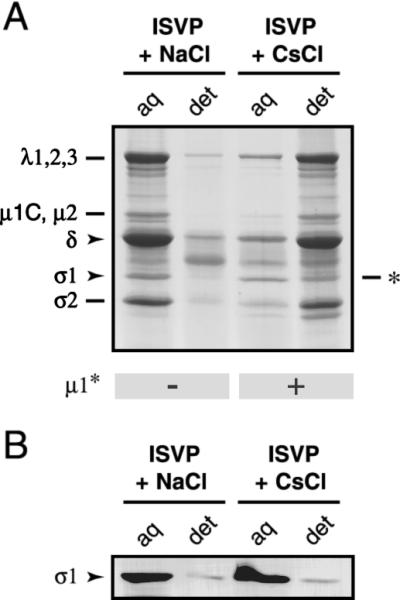
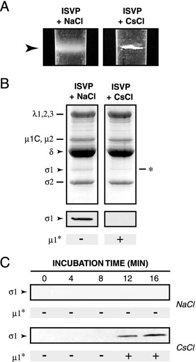
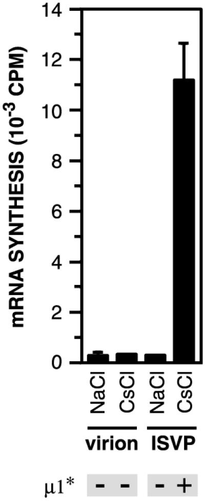
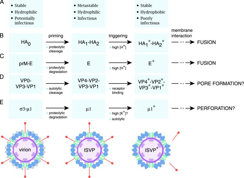
The mechanisms employed by nonenveloped animal viruses to penetrate the membranes of their host cells remain enigmatic. Membrane penetration by the nonenveloped mammalian reoviruses is believed to deliver a partially uncoated, but still large ( approximately 70-nm), particle with active transcriptases for viral mRNA synthesis directly into the cytoplasm. This process is likely initiated by a particle form that resembles infectious subvirion particles (ISVPs), disassembly intermediates produced from virions by proteolytic uncoating. Consistent with that idea, ISVPs, but not virions, can induce disruption of membranes in vitro. Both activities ascribed to ISVP-like particles, membrane disruption in vitro and membrane penetration within cells, are linked to N-myristoylated outer-capsid protein micro 1, present in 600 copies at the surfaces of ISVPs. To understand how micro 1 fulfills its role as the reovirus penetration protein, we monitored changes in ISVPs during the permeabilization of red blood cells induced by these particles. Hemolysis was preceded by a major structural transition in ISVPs, characterized by conformational change in micro 1 and elution of fibrous attachment protein sigma 1. The altered conformer of micro 1 was required for hemolysis and was markedly hydrophobic. The structural transition in ISVPs was further accompanied by derepression of genome-dependent mRNA synthesis by the particle-associated transcriptases. We propose a model for reovirus entry in which (i) primed and triggered conformational changes, analogous to those in enveloped-virus fusion proteins, generate a hydrophobic micro 1 conformer capable of inserting into and disrupting cell membranes and (ii) activation of the viral particles for membrane interaction and mRNA synthesis are concurrent events. Reoviruses provide an opportune system for defining the molecular details of membrane penetration by a large nonenveloped animal virus.


Figures

FIG. 1.
FIG. 1.
Reovirus particles and their capacities to mediate hemolysis. (A) Surface views of the reovirus virion, ISVP, and core obtained from transmission cryoelectron microscopy and three-dimensional image reconstruction as previously reported (22). Color coding of the capsid proteins was applied for clarity (50). Capsid proteins are labeled in a representative fashion. Bar, 20 nm. (B) Purified T3D virions, ISVPs, or cores (1013 particles/ml) or an equal volume of virion buffer lacking particles was incubated with bovine calf RBCs in virion buffer for 15 min on ice or at 32°C. The amount of hemolysis induced by each particle type is shown as the average ± standard deviation from three trials.
FIG. 2.
FIG. 2.
Conformational state of the μ1 protein within hemolysis reactions and reassortant genetic analysis of the difference in hemolysis between T1L and T3D ISVPs. (A) Nonpurified ISVPs of strains T1L (circles), T3D (squares), E3 (diamonds), or EB144 (triangles) (8 × 10¹² particles/ml) were incubated with RBCs in virion buffer for different times at 32°C. Only 0- and 20-min time points were taken for EB144 and E3 ISVPs. Each sample was divided into two equal aliquots. One aliquot was used to measure the amount of hemolysis. Results from two trials are shown. (B) The second aliquot was treated with trypsin for 30 min on ice, and digestion was stopped by addition of soybean trypsin inhibitor. Samples were subjected to SDS-PAGE, and δ, the major fragment of μ1 within ISVPs, was visualized by immunoblotting with μ1-specific monoclonal antibody 10H2. The positions of δ and an unidentified proteolytic fragment derived from δ (∗) are marked. (C) Reassortant strains chosen for this study represent all 16 possible combinations of T1L (L) and T3D (D) alleles for genome segments L2, M2, S1, and S4, which encode outer-capsid proteins λ2, μ1, σ1, and σ3, respectively. Strains are listed in order of decreasing amount of hemolysis. The allelic origin of genome segment M2 is in boldface. For determining the strain behaviors, nonpurified ISVPs (1013 particles/ml) were incubated with RBCs in virion buffer for 7 min at 32°C. Each sample was divided into two equal aliquots. One aliquot was used to measure the amount of hemolysis. Averages from five or six trials (T1L and T3D) and one or two trials (reassortant strains) are shown. The second aliquot was treated with trypsin for 20 min on ice, and digestion was stopped by addition of soybean trypsin inhibitor. Samples were subjected to SDS-PAGE, and the δ fragment of μ1 was visualized by Coomassie staining. Conformational (conf.) change: + or −, loss or retention of δ after trypsin treatment, respectively; nd, not determined.
FIG. 3.
FIG. 3.
Effect of Cs+ ions and RBCs on hemolysis and the conformational state of μ1. (A and B) Nonpurified ISVPs of strains T1L (circles) or T3D (squares) (4 × 10¹² particles/ml) were incubated with RBCs in reaction buffer (50 mM Tris-Cl [pH 7.5]) containing CsCl (300 mM) for different times at 32°C. Measurement of the amount of hemolysis (A) and assessment of the protease sensitivity of δ (B) were carried out as described for Fig. 2A and B. Results from two trials are shown in panel A. (C) Samples were generated as described for panels A and B except that no RBCs were added. Samples were treated with trypsin for 30 min on ice, and digestion was stopped by addition of soybean trypsin inhibitor. Samples were subjected to SDS-PAGE, and the viral proteins were visualized by Coomassie staining.
FIG. 4.
FIG. 4.
Requirement of μ1 conformational change for hemolysis. (A) Schematic diagram for the experiment. Nonpurified T1L ISVPs (4 × 10¹² particles/ml) were incubated in reaction buffer containing CsCl (300 mM) for different times at 32°C. After a 5-min incubation on ice, RBCs were added to each sample, and hemolysis reactions were allowed to proceed for 15 min on ice. (B) The amount of hemolysis in each sample was determined. Results from two trials are shown. (C) Same as panel B except that trypsin was added to samples instead of RBCs. Protease digestion was allowed to proceed for 60 min on ice and was stopped by addition of soybean trypsin inhibitor. Samples were subjected to SDS-PAGE, and the viral proteins were visualized by Coomassie staining.
FIG. 5.
FIG. 5.
Requirement of a protease-sensitive μ1 conformer for hemolysis. (A) Schematic diagram for the experiment. Nonpurified T1L ISVPs (4 × 10¹² particles/ml) were incubated in reaction buffer containing CsCl (300 mM) for 8 min at 32°C and then removed to ice. The sample was divided into four aliquots, and each was treated as indicated. ISVP∗, particle type derived from ISVPs that have undergone μ1 conformational change (see text for more information); try, trypsin; sbti, soybean trypsin inhibitor; try†, trypsin (1 mg/ml) pretreated with soybean trypsin inhibitor (3 mg/ml) for 20 min on ice; rbc†, RBCs pretreated first with trypsin for 20 min on ice and then with soybean trypsin inhibitor for 10 min. (B) The amount of hemolysis in each sample was determined. Averages ± standard deviations from three trials are shown. (C) Same as panel B except that Laemmli sample buffer was added instead of RBCs. Samples were boiled and subjected to SDS-PAGE, and the viral proteins were visualized by Coomassie staining.
FIG. 6.
FIG. 6.
Effect of μ1 conformational change on bis-ANS fluorescence. (A) Nonpurified T1L ISVPs (4 × 10¹² particles/ml) were incubated in reaction buffer containing bis-ANS (25 μm) and NaCl (squares) or CsCl (circles) (300 mM) for different times at 32°C. The amount of bis-ANS fluorescence was then measured. Results from two trials are shown. The conformational state of μ1 in each sample was assessed by trypsin treatment and is indicated below the graph. + and −, loss and retention of δ after trypsin treatment, respectively. μ1∗, protease-sensitive conformer of μ1. (B) Schematic diagram for the order-of-incubation experiment. The experiment was performed as for Fig. 5 except that bis-ANS (25 μM) was added to samples instead of RBCs. (C) The amount of bis-ANS fluorescence in each sample from panel B was determined. Results from two trials are shown as superimposed open and filled bars.
FIG. 7.
FIG. 7.
Effect of μ1 conformational change on Triton X-114 partitioning of viral proteins. Nonpurified T1L ISVPs (4 × 10¹¹ particles/ml) were incubated in reaction buffer containing NaCl or CsCl (200 mM) for 40 min at 37°C and then removed to ice. Samples were extracted with Triton X-114 as described in Materials and Methods. The detergent-poor (aq) and detergent-rich (det) fractions were subjected to SDS-PAGE, followed by Coomassie staining to visualize the viral proteins (A) or immunoblot analysis with a T1L σ1-specific polyclonal antiserum (B). ∗, position of an unidentified proteolytic fragment that comigrated with σ1. The conformational state of μ1 in each fraction was assessed by trypsin treatment and is indicated below the gel.
FIG. 8.
FIG. 8.
Effect of μ1 conformational change on particle association of protein σ1. (A) Nonpurified T1L ISVPs (4 × 10¹² particles/ml) were incubated in reaction buffer containing NaCl or CsCl (300 mM) for 20 min at 32°C and then removed to ice. Each sample was overlaid onto a preformed CsCl gradient (ρ = 1.30 to 1.45 g/cm3, 3.2 ml) and subjected to centrifugation in a Beckman SW60 rotor (30,000 rpm for 2 h at 5°C). Gradients were photographed with a Nikon Coolpix digital camera. Images were cropped and resized in Photoshop, version 5.5 (Adobe Systems, San Jose, Calif.). Arrowhead, position of the band in each gradient. (B) The banded material in panel A was concentrated by TCA precipitation and subjected to SDS-PAGE, followed by Coomassie staining to visualize the viral proteins (top) or immunoblot analysis to visualize σ1. ∗, position of an unidentified proteolytic fragment that comigrated with σ1. The conformational state of μ1 in each sample was assessed by trypsin treatment and is indicated below the gels and immunoblots. (C) Nonpurified T1L ISVPs (4 × 10¹² particles/ml) were incubated in reaction buffer containing NaCl or CsCl (300 mM) for different times at 32°C and then removed to ice. Each sample was overlaid onto a sucrose cushion (20% [wt/vol], 500 μl) and subjected to centrifugation in a Beckman TLA 100.2 rotor (90,000 rpm for 1 h at 5°C). A 200-μl fraction was removed from the top of each gradient, and the proteins in this fraction were concentrated by TCA precipitation and subjected to SDS-PAGE. Protein σ1 was visualized by immunoblot analysis. The conformational state of μ1 in each sample was assessed by trypsin treatment and is indicated below each immunoblot.
FIG. 9.
FIG. 9.
Effect on μ1 conformational change on activity of the particle-associated viral transcriptases. Purified T1L virions and nonpurified T1L ISVPs (4 × 10¹² particles/ml) were incubated in reaction buffer containing NaCl or CsCl (300 mM) at 32°C for 20 min and then removed to ice. Samples were then incubated with a transcription reaction mixture including ribonucleoside triphosphates and [α-32P]GTP for 1 h at 37°C as described previously (44). The amount of 32P incorporated into reovirus mRNA was measured by TCA precipitation followed by liquid scintillation counting (44). Averages ± standard deviations of three trials are shown. The conformational state of μ1 in each sample was assessed by trypsin treatment and is indicated below the graph.
FIG. 10.
FIG. 10.
Parallels between the membrane penetration machines of enveloped and nonenveloped viruses. It is proposed that the membrane penetration protein of the nonenveloped reoviruses (E) resembles the membrane fusion proteins of enveloped viruses such as influenza virus (B) and TBEV (C) as well as the membrane penetration proteins of other nonenveloped viruses such as poliovirus (D). All of these proteins appear to undergo primed and triggered rearrangements that yield a hydrophobic protein conformer capable of interacting with membranes. Protein-membrane interactions have different consequences for the enveloped and nonenveloped viruses: membrane fusion for the former and either membrane pore formation or membrane perforation for the latter. Some essential structural and functional properties of viral particles containing each type of protein conformer are summarized in panel A. (E) Schematic of radial sections of reovirus particle forms containing the different conformers of membrane penetration protein μ1. Also indicated are other changes that accompany the structural transitions in reovirus particles: σ3 degradation (virion to ISVP) and σ1 elution and λ2 conformational change (ISVP to ISVP*). Proteins are colored as in Fig. 1A.

References

    1. Baer, G. S., and T. S. Dermody. 1997. Mutations in reovirus outer-capsid protein σ3 selected during persistent infections of L cells confer resistance to protease inhibitor E64. J. Virol. 71:4921-4928. - PMC - PubMed
    1. Baer, G. S., D. H. Ebert, C. J. Chung, A. H. Erickson, and T. S. Dermody. 1999. Mutant cells selected during persistent reovirus infection do not express mature cathepsin L and do not support reovirus disassembly. J. Virol. 73:9532-9543. - PMC - PubMed
    1. Banerjee, A. K., and A. J. Shatkin. 1970. Transcription in vitro by reovirus-associated ribonucleic acid-dependent polymerase. J. Virol. 6:1-11. - PMC - PubMed
    1. Barton, E. S., J. C. Forrest, J. L. Connolly, J. D. Chappell, Y. Liu, F. J. Schnell, A. Nusrat, C. A. Parkos, and T. S. Dermody. 2001. Junction adhesion molecule is a receptor for reovirus. Cell 104:441-451. - PubMed
    1. Belnap, D. M., D. J. Filman, B. L. Trus, N. Cheng, F. P. Booy, J. F. Conway, S. Curry, C. N. Hiremath, S. K. Tsang, A. C. Steven, and J. M. Hogle. 2000. Molecular tectonic model of virus structural transitions: the putative cell entry states of poliovirus. J. Virol. 74:1342-1354. - PMC - PubMed

Publication types

MeSH terms

LinkOut - more resources