Review
. 2002 Sep;66(3):460-85, table of contents.
doi: 10.1128/MMBR.66.3.460-485.2002.

Evolutionary conservation of reactions in translation

Affiliations
Review

Evolutionary conservation of reactions in translation

M Clelia Ganoza et al. Microbiol Mol Biol Rev. 2002 Sep.

Abstract

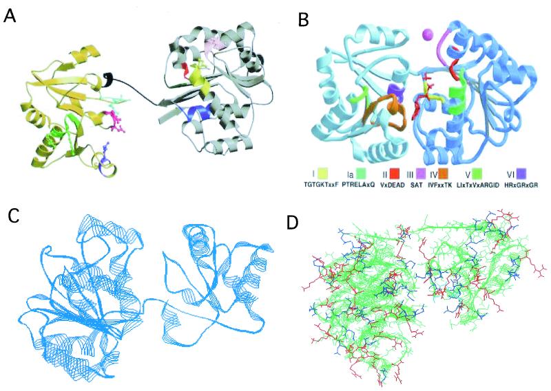
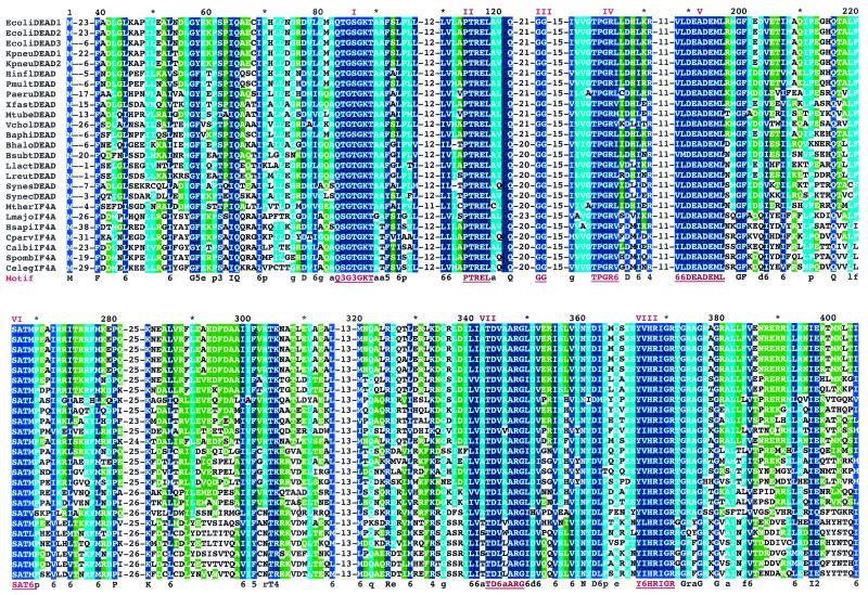
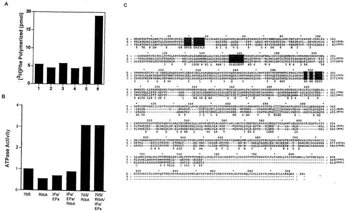
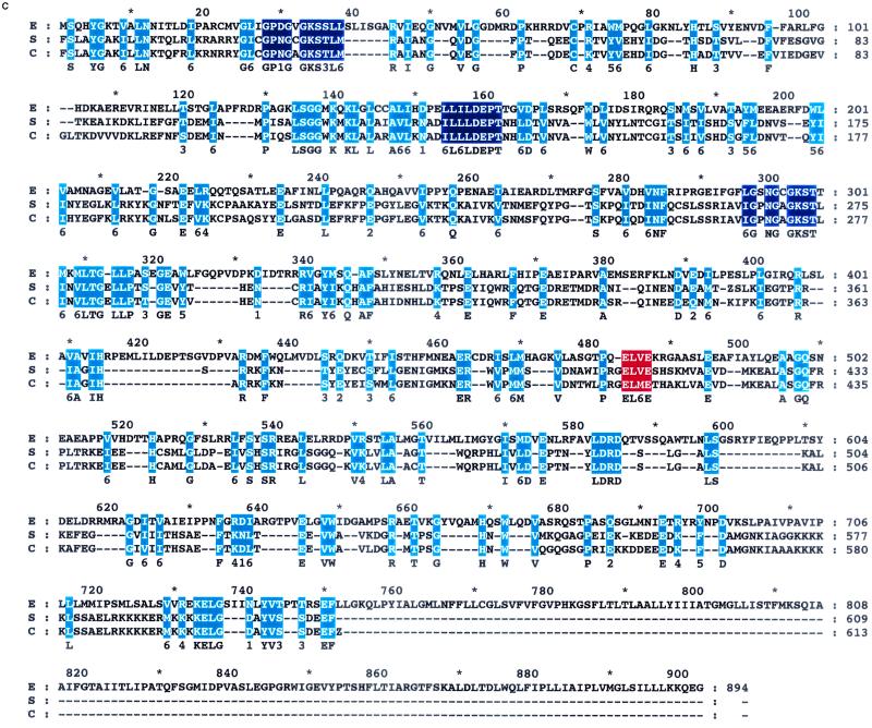
Current X-ray diffraction and cryoelectron microscopic data of ribosomes of eubacteria have shed considerable light on the molecular mechanisms of translation. Structural studies of the protein factors that activate ribosomes also point to many common features in the primary sequence and tertiary structure of these proteins. The reconstitution of the complex apparatus of translation has also revealed new information important to the mechanisms. Surprisingly, the latter approach has uncovered a number of proteins whose sequence and/or structure and function are conserved in all cells, indicating that the mechanisms are indeed conserved. The possible mechanisms of a new initiation factor and two elongation factors are discussed in this context.


Figures

FIG. 1.
FIG. 1.
(A) Ribbon representation of the structure of the full-length eIF4A. The amino-terminal end is shown in brown, and the carboxyl-terminal end is shown in gold. The connecting flexible II residue linker is shown in black. The following domains are colored as indicated: the N-terminal domain, which harbors motif I, which has the Walker A motif, ASQSGTGKT (residues 65 to 72), blue; the Ia motif, PTRELA (residues 97 to 102), yellow; the GG motif (residues 125 to 126), orange; the TPGR (residues 145 to 148), pink; motif II, which harbors the Walker B DEAD motif (residues 169 to 172), red; and motif III, which harbors SAT (residues 200 to 202), green; the C-terminal motif IV, VIFCNTRR (residues 263 to 270), green; the conserved R motif, arginine-298, purple; the RGID motif in motif V (residues 321 to 324), magenta; and the HRIGRGGR (residues 345 to 352) of motif VI, cyan. (Reprinted from reference 25 with permission of the publisher.) (B) Ribbon representation of the structure of the full-length aIF4A from the archaebacterial M. jannaschii derived from the X-ray diffraction data at 1.8-Å resolution. The C-terminal end of the molecule is shown to the left of the figure. The color designation of domains I to IV is given below the figure. (Reprinted from reference 186 with permission of the publisher.) (C) Ribbon representation of the computer-derived structure of the eubacterial IF4A (W2) protein. Approximately 10% of the amino acid sequence of the N terminus of the protein is missing. The E. coli K-12 protein was patterned after the archaebacterial structure in panel B. The C-terminal end of the molecule is shown to the left of the figure. (D) Surface representation of aIF4A of M. jannaschii with the relative distribution of charged amino acid residues. Positively charged residues are shown in blue, and negatively charged residues are shown in red. The molecule is shown with the N-terminal domain to the left and the C-terminal domain to the right. The figure shows that the C-terminal end of the molecule and the bottom of the N-terminal end and the linker that joins the two domains are basic. This figure was generated by the Swiss Plot program.
FIG. 2.
FIG. 2.
Alignment of amino acid sequence of the conserved domains of eIF4A and IF4A (W2) proteins from representative species of eubacteria and archaebacteria. Identical residues within the conserved domains are shown in blue, and highly conserved residues are shown in light blue and green. The sequences were aligned using the Swiss Plot program.
FIG. 3.
FIG. 3.
(A) Secondary structure of the initiation site of the MS2 RNA coat protein cistron, sequence of the start site of the 200-bp unstructured mRNAs, and sequence of the start site of the unstructured 20-bp mRNA. (B) Ribosome-mRNA complexes with unstructured mRNAs or with the structured MS2 RNA. (A) W2 binds unstructured mRNAs (lanes 1 and 2, 20 and 200 bp of [32P]mRNA; lanes 4 and 5, 20 and 200 bp of [32P]mRNA plus 0.2 μg of W2). (B) Binding of 200 bp of unstructured [32P]mRNA to ribosomes with 0, 0.1, 0.2, and 0.4 μg of W2 (lanes 1 to 4). (C) Ternary initiation complexes, f[35S]Met-tRNA-ribosome-MS2 RNA, require W2. (D) Ternary initiation complexes, fMet[35S]Met-tRNA-ribosome-unstructured mRNA, do not require W2 (lane 1, no mRNA; lane 2, 200 bp of mRNA; lane 3, 200 bp of mRNA plus 0.1 μg of W2; lane 4, 20 bp of mRNA; lane 5, 20 bp of mRNA plus 0.1 μg of W2). See reference 122 for experimental details.
FIG. 4.
FIG. 4.
Alignment of amino acid sequences of the EFP proteins from eubacterial, archaebacterial, and eukaryotic sources. The Lys-31 motif in the eubacterial sequences bears an unusual modification. Lys-54 of the eukaryotic sequences is modified by hypusine. Identical residues in the eukaryotic sequences are shown in yellow, sequences unique to eubacterial sequences are shown in blue, and conserved residues are shown in green.
FIG. 5.
FIG. 5.
(A) Ribbon diagram and representative sketch of the structure of aIF5A from M. jannaschii. The ribbon structure was derived from X-ray diffraction patterns at 1.8-Å resolution. The sketch (A) shows the eleven β sheets in two domains of the molecule joined by flexible links that contain the Lys-54 site of hypusine modification. (Reprinted from reference 108 with permission of the publisher.) (B) Ribbon structure of the eIF5A, showing the two domains of the molecule linked by the flexible linker that harbors the hypusine residue. The molecule bears opposite charges in the C- and N-terminal ends.
FIG. 6.
FIG. 6.
(A) Requirement for EFP and L16 in peptide bond synthesis by reconstituted 70S ribosomes (○, no EFP; ▪, with 2 μg of EFP); the cores were reconstituted with L16 (▴), and then EFP was added (•). (B) Synthesis of dipeptides from fMet-tRNA and 5′ CCA amino acids; association constants in the presence or absence of EFP as a function of the length of the amino acid side chain. Data are from references 52 and 64. The sizes of the amino acid side chains are derived from X-ray diffraction data.
FIG. 7.
FIG. 7.
Requirement for RbbA and EFP in MS2 programmed synthesis reconstituted from homogeneous proteins. Data are from reference .
FIG. 8.
FIG. 8.
Hybrid state model for the translational elongation cycle (128). tRNA-binding sites on the 50S and 30S subunits are represented by the upper and lower rectangles, respectively. The 50S subunit harbors A (aminoacyl), P (peptidyl), and E (exit) sites, and the 30S subunit has only A and P sites. tRNAs are represented by vertical bars, and amino acids are represented by small circles. mRNA is represented as a line bound to the 30S subunit. The directional movement of the acylated and deacylated tRNAs through the ribosome, catalyzed by elongation factors EFTu and EFG, during the translational cycle is indicated, as are the proposed stimulation of the peptidyl transferase by EFP and tRNA ejection by RbbA. PT, peptidyl transfer.
FIG. 9.
FIG. 9.
(A) Requirement for RbbA in synthesis with pure elongation factors. Lane 1, EFTu, EFG, GTP (EF); lane 2, EF plus AMPPcP; lane 3, EF plus ATP; lane 4, EF plus RbbA; lane 5, EF plus RbbA plus AMPPcP; lane 6, EF plus RbbA plus ATP. (B) Ribosome-dependent ATPase activity of RbbA. Data are from references 105 to 107. (C) Predicted amino acid sequence of RbbA. The predicted amino acid sequence of RbbA based on open reading frame yhih sequenced by Blattner et al. (15) is shown as blue-highlighted letters representing amino acids that are part of the ABC. Blue-highlighted letters represent amino acids that make up the aminoacyl-tRNA synthetase motif shared with the yeast EF3. Yellow-highlighted letters represent amino acids that are identical or very similar in the S. cerevisiae (S), C. albicans EF3 (C), and E. coli (E) RbbA proteins. The sequences were aligned with the Swiss Plot program.
FIG. 9.
FIG. 9.
(A) Requirement for RbbA in synthesis with pure elongation factors. Lane 1, EFTu, EFG, GTP (EF); lane 2, EF plus AMPPcP; lane 3, EF plus ATP; lane 4, EF plus RbbA; lane 5, EF plus RbbA plus AMPPcP; lane 6, EF plus RbbA plus ATP. (B) Ribosome-dependent ATPase activity of RbbA. Data are from references 105 to 107. (C) Predicted amino acid sequence of RbbA. The predicted amino acid sequence of RbbA based on open reading frame yhih sequenced by Blattner et al. (15) is shown as blue-highlighted letters representing amino acids that are part of the ABC. Blue-highlighted letters represent amino acids that make up the aminoacyl-tRNA synthetase motif shared with the yeast EF3. Yellow-highlighted letters represent amino acids that are identical or very similar in the S. cerevisiae (S), C. albicans EF3 (C), and E. coli (E) RbbA proteins. The sequences were aligned with the Swiss Plot program.

References

    1. Adhin, M. R., and J. van Duin. 1990. Scanning model for translational reinitiation in eubacteria. J. Mol. Biol. 213:811-818. - PubMed
    1. Ævarsson, A., E. Brazhnikov, M. Garber, J. Zheltonosova, Y. Chirgadze, S. al-Karadaghi, L. A. Svensson, and A. Liljas. 1994. Three-dimensional structure of the ribosomal translocase: elongation factor G from Thermus thermophilus. EMBO J. 13:3669-3677. - PMC - PubMed
    1. Ames, G. F., C. S. Mimura, S. R. Holbrook, and V. Shyamala. 1992. Traffic ATPases: a superfamily of transport proteins operating from Escherichia coli to humans. Adv. Enzymol. Relat. Areas Mol. Biol. 65:1-47. - PubMed
    1. An, G., B. R. Glick, J. D. Friesen, and M. C. Ganoza. 1980. Identification and quantification of elongation factor EFP in E. coli. Can. J. Biochem. 97:23-28.
    1. Aoki, H., S. L. Adams, D. Watson, M. Yaguchi, I. Kozieradzki, and M. C. Ganoza. 1997. Molecular characterization of the efp gene product involved in a peptidyl transferase reaction. Biochimie 79:7-11. - PubMed

Publication types

MeSH terms

LinkOut - more resources