. 2002 Sep 17;99(19):12269-74.
doi: 10.1073/pnas.192445899. Epub 2002 Sep 3.

MYO18B, a candidate tumor suppressor gene at chromosome 22q12.1, deleted, mutated, and methylated in human lung cancer

Affiliations

MYO18B, a candidate tumor suppressor gene at chromosome 22q12.1, deleted, mutated, and methylated in human lung cancer

Michiho Nishioka et al. Proc Natl Acad Sci U S A. .

Abstract

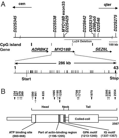
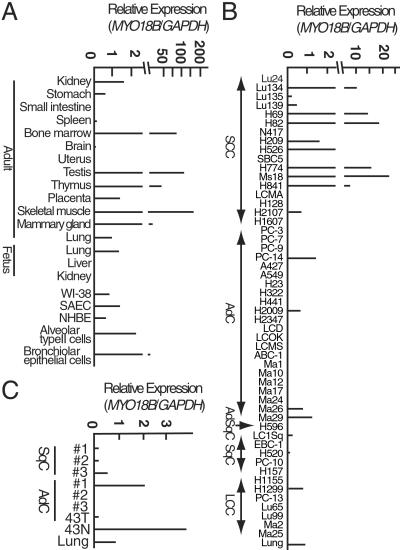
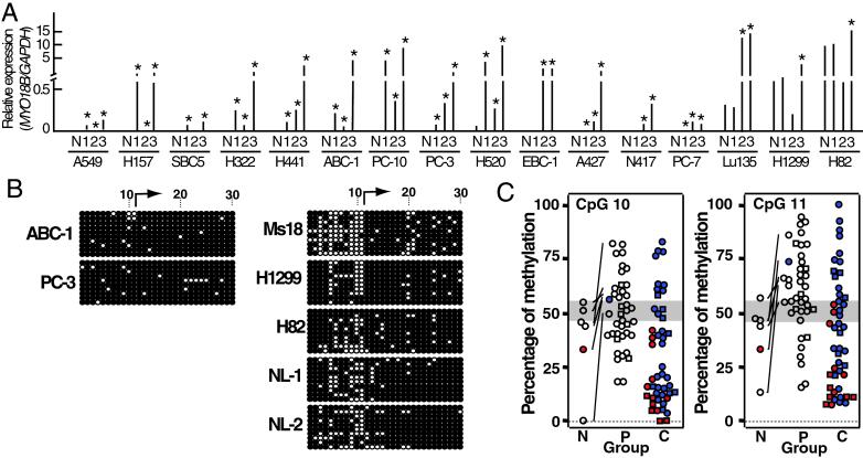
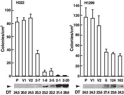
Loss of heterozygosity on chromosome 22q has been detected in approximately 60% of advanced nonsmall cell lung carcinoma (NSCLC) as well as small cell lung carcinoma (SCLC), suggesting the presence of a tumor suppressor gene on 22q that is involved in lung cancer progression. Here, we isolated a myosin family gene, MYO18B, located at chromosome 22q12.1 and found that it is frequently deleted, mutated, and hypermethylated in lung cancers. Somatic MYO18B mutations were detected in 19% (14/75) of lung cancer cell lines and 13% (6/46) of primary lung cancers of both SCLC and NSCLC types. MYO18B expression was reduced in 88% (30/34) of NSCLC and 47% (8/17) of SCLC cell lines. Its expression was restored by treatment with 5-aza-2'-deoxycytidine in 11 of 14 cell lines with reduced MYO18B expression, and the promoter CpG island of the MYO18B gene was methylated in 17% (8/47) of lung cancer cell lines and 35% (14/40) of primary lung cancers. Furthermore, restoration of MYO18B expression in lung carcinoma cells suppressed anchorage-independent growth. These results indicate that the MYO18B gene is a strong candidate for a novel tumor suppressor gene whose inactivation is involved in lung cancer progression.


Figures

Fig 1.
Fig 1.
Structures of the MYO18B gene and MYO18B protein. (A) Physical and transcriptional maps of the region containing the MYO18B gene. STS markers are marked on the top. Locations of CpG islands and three genes are indicated. The region of homozygous deletion in a SCLC cell line, Lu24, is indicated by a dashed horizontal line. Exon-intron organization of the MYO18B gene is depicted as vertical bars. Predicted transcriptional start and stop sites are indicated by vertical bars under exons 2 and 43, respectively. (B) Schematic diagram of a domain structure of MYO18B protein and positions of mutations in lung cancer cells. Numbers on the top indicate codon numbers. Closed arrowheads, somatic mutations; open arrowheads, base substitutions detected only in cell lines but not in 50 unrelated individuals. Putative functional domains are indicated at the bottom.
Fig 2.
Fig 2.
Expression of the MYO18B gene. SCC, small cell carcinoma; AdC, adenocarcinoma; AdSqC, adenosquamous carcinoma; SqC, squamous cell carcinoma; LCC, large cell carcinoma. (A and C) Expression in normal human tissues and cancerous and noncancerous lung cells. Noncancerous tissue (43N) and cancerous tissue (43T) were obtained from the same patient. (B) Expression in lung cancer cell lines.
Fig 3.
Fig 3.
Expression and methylation of the MYO18B gene in cancerous and noncancerous lung cells. (A) Restoration of MYO18B expression after treatment with 5-aza-dC and/or TSA in 14 cell lines with reduced (or without) MYO18B expression. N, no treatment; 1, 5-aza-dC treatment; 2, TSA treatment; 3, 5-aza-dC and TSA treatment. Each result is the average of two independent tests. * indicates expression levels were more than twice as much as basal levels. (B) Bisulfite sequencing of the 5′ CpG-rich region of the MYO18B gene in lung cancer cell lines without (ABC-1 and PC-3) or with (Ms18, H1299, and H82) MYO18B expression and two normal lung tissues (NL-1 and NL-2). Each circle indicates a CpG site in the primary DNA sequence, and each line of circles represents analysis of a single cloned allele. The numbers of the CpG site are indicated at the top and are identical to the numbers in Fig. 5. ○, unmethylated CpG sites; •, methylated CpG sites; arrow, transcription start site. (C) Methylation status at CpG 10/11 in normal lung tissues and lung cancer cells. The ranges of 50% methylation (n = 7, mean ± 3 SD) at CpG 10/11 are shaded. Paired cancerous and noncancerous lung tissues are connected with solid lines. Samples with MYO18B mutation, square; samples with or with reduced (or without) MYO18B expression, filled in red or blue, respectively. Open squares and circles indicate samples in which MYO18B expression was not determined. Groups N, P, and C indicate normal lung tissue (n = 6), primary tumor (n = 40), and cell line (n = 47), respectively.
Fig 4.
Fig 4.
Suppression of proliferation and anchorage-independent growth by exogenous MYO18B expression. H322 (P), vector-transfected controls (V1, V2), and MYO18B-transfected clones (, , , , 2–20) were analyzed for population doubling time and colony formation in soft agar (Left). H1299 (P), vector-transfected controls (V1, V2), and MYO18B-transfected clones (, 124, 163) were also analyzed (Right). The arrowhead indicates the expression of FLAG-tagged MYO18B protein (285 kDa) by Western blot analysis. Twenty micrograms of whole-cell lysate was loaded on each lane. The doubling time [DT (hr)] was calculated from the logarithmic phase of the growth curve. The ability to form colonies in soft agar was measured by counting the number of visible colonies/cm2 in triplicate of a 6-well plate. Error bars indicate SD.

References

    1. Shiseki M., Kohno, T., Nishikawa, R., Sameshima, Y., Mizoguchi, H. & Yokota, J. (1994) Cancer Res. 54, 5643-5648. - PubMed
    1. Kawanishi M., Kohno, T., Otsuka, T., Adachi, J., Sone, S., Noguchi, M., Hirohashi, S. & Yokota, J. (1997) Carcinogenesis 18, 2057-2062. - PubMed
    1. Virmani A. K., Fong, K. M., Kodagoda, D., McIntire, D., Hung, J., Tonk, V., Minna, J. D. & Gazdar, A. F. (1998) Genes Chromosomes Cancer 21, 308-319. - PubMed
    1. Testa J. R., Siegfried, J. M., Liu, Z., Hunt, J. D., Feder, M. M., Litwin, S., Zhou, J. Y., Taguchi, T. & Keller, S. M. (1994) Genes Chromosomes Cancer 11, 178-194. - PubMed
    1. Mertens F., Johansson, B., Hoglund, M. & Mitelman, F. (1997) Cancer Res. 57, 2765-2780. - PubMed

Publication types