. 2002 Sep;161(3):831-9.
doi: 10.1016/S0002-9440(10)64243-5.

The lack of thrombospondin-1 (TSP1) dictates the course of wound healing in double-TSP1/TSP2-null mice

Affiliations

The lack of thrombospondin-1 (TSP1) dictates the course of wound healing in double-TSP1/TSP2-null mice

Azin Agah et al. Am J Pathol. 2002 Sep.

Abstract

Thrombospondin (TSP) 1 and 2, share the same overall structure and interact with a number of the same cell-surface receptors. In an attempt to elucidate their biological roles more clearly, we generated double-TSP1/TSP2-null animals and compared their phenotype to those of TSP1- and TSP2-null mice. Double-null mice exhibited an apparent phenotype that primarily represented the sum of the abnormalities observed in the single-null mice. However, surprisingly, the wound-healing response in double-null mice resembled that in TSP1-null animals and differed from that in TSP2-nulls. Thus, although the excisional wounds of TSP2-null mice are characterized by increased neovascularization and heal at an accelerated rate, TSP1-null and double-null animals demonstrated delayed healing, as indicated by the prolonged persistence of inflammation and delayed scab loss. Immunohistochemical analysis showed that, similar to TSP1-null mice, the granulation tissue of double-null mice was not excessively vascularized. Furthermore as in TSP1-nulls, decreases in macrophage recruitment and in the levels of monocyte chemoattractant protein-1 indicated that the inflammatory phase of the wound-healing response was impaired in double-null mice. Our data demonstrate that the consequences of a lack of TSP1 predominate in the response of double-null mice, and dictate the course of wound healing. These findings reflect distinct temporal and spatial expressions of TSP1 and TSP2 in the healing wound.


Figures

Figure 1.
Figure 1.
Genotyping and protein analyses. A: Southern analysis of tail DNA from TSP1−/−/TSP2+/+ (lane 1), double-TSP1−/−/TSP2−/− (lane 2), wild-type (lane 3), and TSP1+/+/TSP2−/− animals (lane 4). The probe detects 5.8-kb and 4.4-kb fragments in StuI digests of the wild-type and targeted TSP1 alleles, respectively. B: Southern analysis of tail DNA from TSP1−/−/TSP2+/− (lane 1), TSP1+/+/TSP2−/− (lane 2), and double-TSP1−/−/TSP2−/− animals (lane 3). The probe detects 6.0-kb and 4.8-kb fragments in BamHI digests of the wild-type and targeted TSP2 alleles, respectively. C: PCR analysis of tail DNA from wild-type (lane 1), TSP1+/+/TSP2−/− (lane 2), double-TSP1−/−/TSP2−/− (lane 3), and TSP1−/−/TSP2+/+ animals (lane 4). The 400-bp band identifies the targeted TSP1 allele and the 700-bp band identifies the wild-type TSP1 allele. Lane M: molecular weight marker. D: PCR analysis of tail DNA from wild-type (lane 1), TSP1−/−/TSP2+/+ (lane 2), double-TSP1−/−/TSP2−/− (lane 3), and TSP1+/+/TSP2−/− animals (lane 4). The 900-bp band identifies the targeted TSP2 allele, and the 536-bp band identifies the wild-type TSP2 allele. Lane M: molecular weight marker. E: Western analysis for TSP1 in conditioned media of mouse dermal fibroblasts. Lane 1, Conditioned media from wild-type cells; lane 2, conditioned media from TSP1−/−/TSP2+/+ cells; lane 3, conditioned media from TSP1+/+/TSP2−/− cells; lane 4, conditioned media from double-TSP1−/−/TSP2−/− cells. F: Western analysis for TSP2 in conditioned media of mouse dermal fibroblasts. Lane 1, Conditioned media from wild-type cells; lane 2, conditioned media from TSP1−/−/TSP2+/+ cells; lane 3, conditioned media from TSP1+/+/TSP2−/− cells; lane 4, conditioned media from double-TSP1−/−/TSP2−/− cells.
Figure 2.
Figure 2.
Histological analysis of lung tissue from wild-type (A, D), TSP2-null (B, E), and double-null (C, F) mice. Tissue sections were stained with H&E (A–C) or with antibodies against Mac-3 and a methyl green counterstain (D–F). Compared with the normal histology observed in H&E-stained sections of wild-type and TSP2-null lungs, lungs of double-null mice displayed abnormalities marked by patchy consolidation associated with acute and chronic pneumonia. Immunostaining with Mac-3 demonstrated prominent macrophage infiltration in lungs of double-TSP1−/−/TSP2−/− mice. Little or no immunoreactivity was detected in lungs of wild-type or TSP2-null mice. Scale bar, 100 μm. WT, wild-type; DKO, double knockout.
Figure 3.
Figure 3.
Collagen fiber patterns in the granulation tissue of healing wounds. Sections of day 14 wounds were visualized with Masson’s trichrome stain. Collagen fiber patterns in wild-type and TSP2-null wounds resemble each other in that fibers are tightly packed, but fibers in wild-type wounds have a parallel orientation to the epidermis. Collagen fiber patterns in TSP1-null and double-TSP1/TSP2-null animals also resemble each other in the appearance of the fiber weave, but differ from those observed in wild-type and TSP2-null mice in being less densely packed. Scale bar, 50 μm. WT, wild-type; KO, knockout; DKO, double knockout.
Figure 4.
Figure 4.
Temporal distribution of TSP1 and TSP2 during healing of excisional wounds. Sections of wounds were stained with antibodies against murine TSP1 and TSP2. A: Time course of expression of TSP1 and TSP2 during healing of excisional wounds. Relative levels of TSP1 and TSP2 were measured by histomorphometry after immunohistochemical analysis. Because the antibodies to murine TSP1 and TSP2 are likely to possess different affinities for their antigens, quantitative comparisons are not possible. B: Temporal distribution of TSP1 in wild-type and TSP2-null wounds. C: Temporal distribution of TSP2 in TSP1-null and wild-type wounds. Values are arbitrary units. Bars indicate the mean ± SD.
Figure 5.
Figure 5.
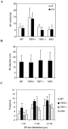
TSP1-null and double-TSP1/TSP2-null animals lack the increased vascularity observed in TSP2-null animals. Sections of days 7 and 14 wounds were stained with anti-PECAM-1 antibodies and visualized by the peroxidase reaction. A total of 16 sections from four animals/genotype/time point were quantified. A: The wound beds in both TSP1-null and double-null wounds lack the increased vascularity observed in TSP2-null animals. B: Blood vessel (BV) sizes were similar in all four groups at day 7. C: Blood vessel size distribution appeared similar in all four groups at day 7. DKO, double knockout. Bars indicate the mean ± SD; *, significantly different from control, P ≤ 0.05.
Figure 6.
Figure 6.
Macrophage infiltration into excisional wounds. A: Paraffin-embedded sections of day 7 wounds were incubated with monoclonal antibodies against murine macrophage-specific antigen (Mac 3) and visualized by the peroxidase reaction. For each section, the number of macrophages was counted in five to eight random high-power fields with the aid of an optical grid. The numbers of macrophages in both TSP1-null and double-TSP1/TSP2-null wounds were significantly reduced. B: MCP-1 protein levels in excisional wounds. The levels of MCP-1 were similar 12 hours after injury, but were significantly lower at days 1 and 3 after injury in both TSP1-null and double-TSP1/TSP2-null wounds. Bars indicate the mean ± SD; *, significantly different from control, P ≤ 0.05. WT, wild-type; DKO, double knockout.
Figure 7.
Figure 7.
TGF-β1 protein levels in day 3 excisional wounds. Levels of total and active TGF-β1 in both TSP1-null and double-TSP1/TSP2-null wounds were significantly reduced compared with levels in wild-type or TSP2-null wounds. Bars indicate the mean ± SD; *, significantly different from control, P ≤ 0.05. WT, wild-type; DKO, double knockout.

References

    1. Frazier WA: Thrombospondins. Curr Opin Cell Biol 1991, 3:792-799 - PubMed
    1. Bornstein P, Armstrong LC, Hankenson KD, Kyriakides TR, Yang Z: Thrombospondin 2, a matricellular protein with diverse functions. Matrix Biol 2000, 19:557-568 - PubMed
    1. Chen H, Herndon ME, Lawler J: The cell biology of thrombospondin-1. Matrix Biol 2000, 19:597-614 - PubMed
    1. Lawler J: The functions of thrombospondin-1 and -2. Curr Opin Cell Biol 2000, 12:634-640 - PubMed
    1. Bornstein P: Thrombospondins as matricellular modulators of cell function. J Clin Invest 2001, 107:929-934 - PMC - PubMed

Publication types