

Susceptibility to human immunodeficiency virus-1 infection of human foreskin and cervical tissue grown in explant culture
- PMID: 12213715
- PMCID: PMC1867269
- DOI: 10.1016/S0002-9440(10)64247-2
Susceptibility to human immunodeficiency virus-1 infection of human foreskin and cervical tissue grown in explant culture
Abstract
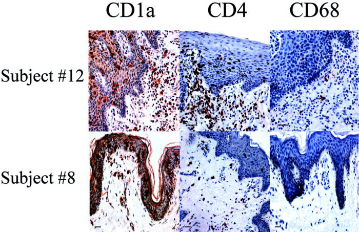
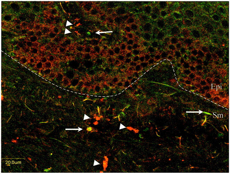
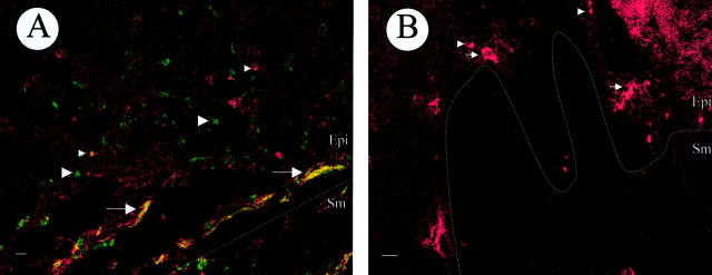
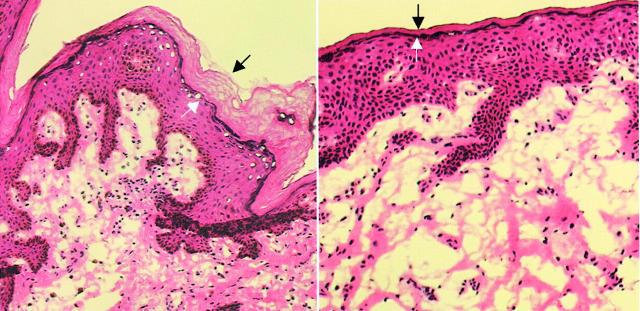
Numerous studies have indicated a protective effect of male circumcision against acquisition of human immunodeficiency virus (HIV)-1. We investigated mechanisms responsible for the possible increased HIV-1 susceptibility of human foreskin. Foreskins from eight pediatric and six adult patients with (n = 3) and without (n = 11) histories of sexually transmitted disease were evaluated. Six cervical biopsies from HIV-1-seronegative women were included as controls. CD4(+) T cells, macrophages, and Langerhans' cells (LCs) were quantified using image analysis. Cells expressing HIV-1 co-receptors CCR5 and CXCR4 were quantified using immunofluorescence and image analysis. Foreskin biopsies were infected ex vivo in organotypic culture with HIV-1. HIV-1 DNA copies in foreskin and cervical mucosal tissue were compared and the infected cell phenotype was determined. Foreskin mucosa contained higher mean proportions of CD4(+) T cells (22.4%), macrophages (2.4%), and LCs (11.5%) in adults than in children (4.9%, 0.3%, and 6.2%, respectively) or in cervical mucosa (6.2%, 1.4%, and 1.5%, respectively). The highest proportions of CD4(+) T cells and LCs occurred in patients with a history of infection. Foreskin immune cells expressed predominantly the CCR5 HIV-1 co-receptor. Adult foreskin mucosa had greater susceptibility to infection with HIV(bal) than cervical mucosa or the external surface of foreskin tissue. Circumcision likely reduces risk of HIV-1 acquisition in men by decreasing HIV-1 target cells.
Figures
References
-
- : Joint United Nations Programme on HIV-1/AIDS (UNAIDS) and World Health Organization (WHO): AIDS Epidemic Update—December 2001 2002. UNAIDS, Geneva
-
- Bailey RC, Plummer FA, Moses S: Male circumcision and HIV prevention: current knowledge and future research directions. Lancet Infect Dis 2002, 1:223-231 - PubMed
-
- Weiss HA, Quigley MA, Hayes RJ: Male circumcision and risk of HIV-1 infection in sub-Saharan Africa: a systematic review and meta-analysis. AIDS 2000, 14:2261-2370 - PubMed
Publication types
MeSH terms
Grants and funding
LinkOut - more resources
Full Text Sources
Medical
Research Materials
