. 2002 Sep 2;158(5):929-40.
doi: 10.1083/jcb.200112081. Epub 2002 Sep 3.

Regulation of membrane fusion by the membrane-proximal coil of the t-SNARE during zippering of SNAREpins

Affiliations

Regulation of membrane fusion by the membrane-proximal coil of the t-SNARE during zippering of SNAREpins

Thomas J Melia et al. J Cell Biol. .

Abstract

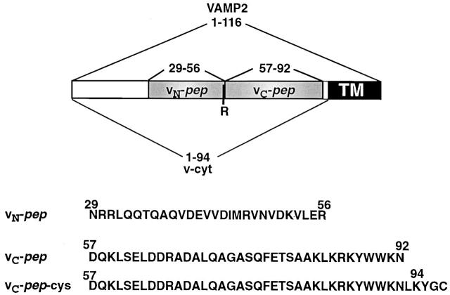
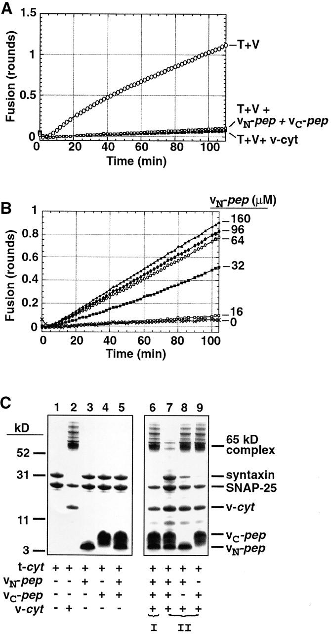
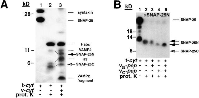
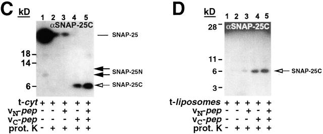
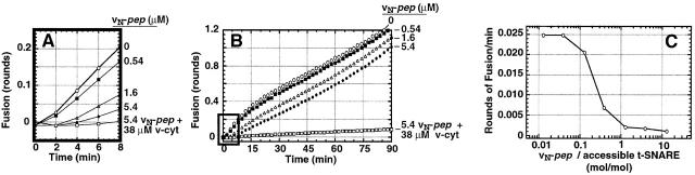
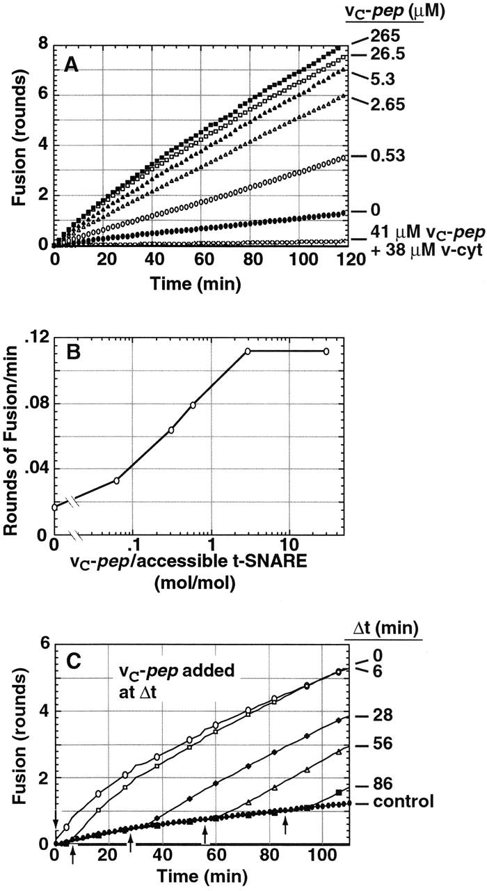
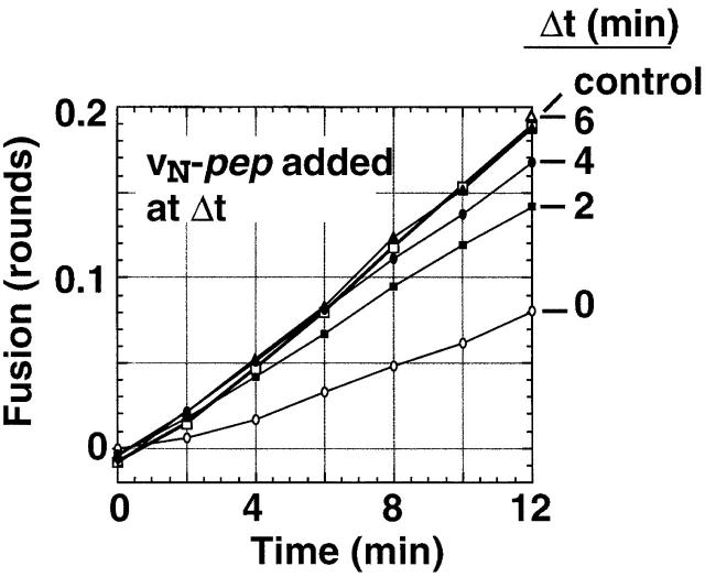
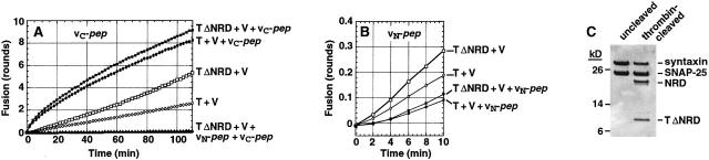
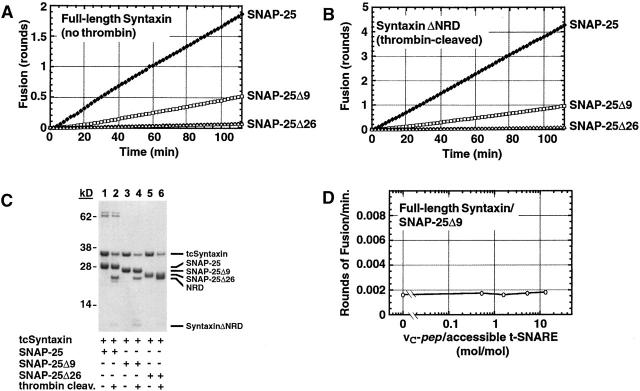
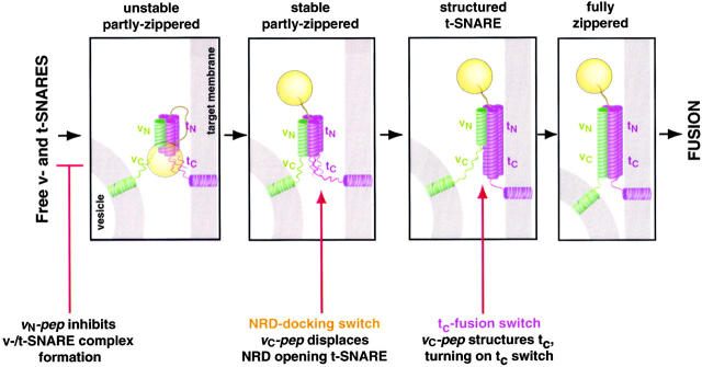
We utilize structurally targeted peptides to identify a "tC fusion switch" inherent to the coil domains of the neuronal t-SNARE that pairs with the cognate v-SNARE. The tC fusion switch is located in the membrane-proximal portion of the t-SNARE and controls the rate at which the helical bundle that forms the SNAREpin can zip up to drive bilayer fusion. When the fusion switch is "off" (the intrinsic state of the t-SNARE), zippering of the helices from their membrane-distal ends is impeded and fusion is slow. When the tC fusion switch is "on," fusion is much faster. The tC fusion switch can be thrown by a peptide that corresponds to the membrane-proximal half of the cognate v-SNARE, and binds reversibly to the cognate region of the t-SNARE. This structures the coil in the membrane-proximal domain of the t-SNARE and accelerates fusion, implying that the intrinsically unstable coil in that region is a natural impediment to the completion of zippering, and thus, fusion. Proteins that stabilize or destabilize one or the other state of the tC fusion switch would exert fine temporal control over the rate of fusion after SNAREs have already partly zippered up.


Figures

Figure 1.
Figure 1.
VAMP2 domain organization and recombinant constructs. The borders of the various constructs are defined by amino acid positions. Together, the synthetic peptides vN-pep and vC-pep (shaded gray) encompass the entire coiled-coil domain of VAMP2 as determined by crystal structure analysis (Sutton et al., 1998). TM, transmembrane region.
Figure 2.
Figure 2.
vN-pep and vC-pep bind the t-SNARE via the functionally relevant VAMP2 binding site. (A) vN-pep and vC-pep, when added together, compete with v-liposomes to inhibit fusion as effectively as v-cyt. Lipid-mixing fusion assays of t- and v-liposomes (T + V) were performed at 37°C as described in Materials and methods. Protein- and peptide-mediated inhibitions were tested by adding buffer (○), 50 μM vN-pep + 38 μM vC-pep (□), or 31 μM v-cyt (▪). (B) The VAMP2 coiled coil can be broken at the zero layer and still function in liposome fusion. vC-pep-cys was covalently modified with a C45 isoprenoid and incorporated into labeled liposomes. vC-pep-cys-C45 liposome fusion is dependent upon the concentration of vN-pep (no vN-pep, X; 16 μM, □; 32 μM, ▪; 64 μM, ○; 96 μM, •; 160 μM, ▵). (C) Together, the two peptides block SNARE complex formation. To test for the ability to form SDS-resistant complexes (indicated by the presence of the 65-kD band), 10 μg t-cyt was incubated with buffer (lane 1), 6 μg v-cyt (lane 2), 10 μg vN-pep (lane 3), 10 μg vC-pep (lane 4), or both peptides (lane 5) for 20 min at RT, and then immediately mixed with buffer without boiling and analyzed by SDS-PAGE and Coomassie blue staining. In order of addition experiments, to test for competition of binding at the VAMP2 binding site (lanes 6–9), t-cyt was preincubated with one component for 20 min before addition of the second component for 20 additional min. (I) t-cyt was incubated with v-cyt, and then both peptides. (II) t-cyt was incubated with one or both peptides, and then v-cyt. (Note: the SNAP-25 that does not become incorporated into the 65-kD SDS-resistant complex is derived from an excess of free SNAP-25 in the t-cyt preparation).
Figure 3.
Figure 3.
vC-pep binding changes the t-SNARE conformation. t-cyt was incubated in the presence or absence of VAMP2 and VAMP2 peptides for 20 min at RT, and then was digested with proteinase K, as described in Materials and methods. Concentrations of the components in the digest were as follows: 2.2 mg/ml t-cyt, 2 mg/ml vC-pep, 2 mg/ml vN-pep, 1.2 mg/ml v-cyt, and 50 μg/ml proteinase K. (A) t-cyt– and t-cyt + v-cyt–digested samples were boiled, resolved by SDS-PAGE on a 12% gel (Novex) with bis-Tris buffer, and stained with Coomassie brilliant blue. The bands were identified by microsequencing and mass spectrometry. The lines between lanes 2 and 3 indicate coils that have gotten smaller (SNAP-25N) or larger (H3 and SNAP-25C). The low molecular mass band originating from v-cyt is a proteinase K–resistant fragment from the highly charged COOH terminus of v-cyt, and is observed when either v-cyt or vC-pep are proteolyzed alone. (B and C) Proteolytic fragments of SNAP-25 were detected by Western blot analysis. Samples were digested and resolved as above and transferred to nitrocellulose. The Western blot was probed, stripped, and reprobed sequentially with antibodies against the SNAP-25 NH2 terminus (B; Cl 71.1) and the SNAP-25 COOH terminus (C; anti–amino acids 207–218). (D) t-liposomes were incubated with or without peptides and proteolyzed as in A–C. Protection of the COOH terminus of SNAP-25 was detected by Western blotting with the same peptide antibody as in C. Note that the large amount of full-length SNAP-25 remaining after proteolysis reflects the fully protected protein in the vesicle lumen.
Figure 3.
Figure 3.
vC-pep binding changes the t-SNARE conformation. t-cyt was incubated in the presence or absence of VAMP2 and VAMP2 peptides for 20 min at RT, and then was digested with proteinase K, as described in Materials and methods. Concentrations of the components in the digest were as follows: 2.2 mg/ml t-cyt, 2 mg/ml vC-pep, 2 mg/ml vN-pep, 1.2 mg/ml v-cyt, and 50 μg/ml proteinase K. (A) t-cyt– and t-cyt + v-cyt–digested samples were boiled, resolved by SDS-PAGE on a 12% gel (Novex) with bis-Tris buffer, and stained with Coomassie brilliant blue. The bands were identified by microsequencing and mass spectrometry. The lines between lanes 2 and 3 indicate coils that have gotten smaller (SNAP-25N) or larger (H3 and SNAP-25C). The low molecular mass band originating from v-cyt is a proteinase K–resistant fragment from the highly charged COOH terminus of v-cyt, and is observed when either v-cyt or vC-pep are proteolyzed alone. (B and C) Proteolytic fragments of SNAP-25 were detected by Western blot analysis. Samples were digested and resolved as above and transferred to nitrocellulose. The Western blot was probed, stripped, and reprobed sequentially with antibodies against the SNAP-25 NH2 terminus (B; Cl 71.1) and the SNAP-25 COOH terminus (C; anti–amino acids 207–218). (D) t-liposomes were incubated with or without peptides and proteolyzed as in A–C. Protection of the COOH terminus of SNAP-25 was detected by Western blotting with the same peptide antibody as in C. Note that the large amount of full-length SNAP-25 remaining after proteolysis reflects the fully protected protein in the vesicle lumen.
Figure 4.
Figure 4.
vN-pep reversibly inhibits fusion. t-liposomes (20 mg protein) were incubated with vN-pep for 7 min at RT before initiating the fusion assay by addition of v-liposomes. (A) vN-pep reduces the initial rate of liposome fusion, almost to zero. Data are plotted in rounds of fusion (Parlati et al., 1999). No peptide (○), 0.54 μM vN-pep (▪), 1.6 μM vN-pep (▵), 5.4 μM vN-pep (•), 5.4 μM vN-pep + 38 μM V-cyt (□). (B) After a prolonged lag-phase, fusion recovers. The initial kinetics in A correspond to the black-boxed region. (C) vN-pep–induced lag is dose dependent and maximal at concentrations that are approximately stoichiometric with accessible t-SNARE. The rate of fusion over the first 6 min of the fusion assay is plotted at varying peptide to t-SNARE ratios in terms of rounds of fusion per minute. Accessible t-SNARE is defined as t-SNAREs in which the coiled-coil domain is facing out on the liposome and is estimated to be 70% of the total t-SNARE (Weber et al., 1998).
Figure 5.
Figure 5.
vC-pep stimulates liposome fusion. t-liposomes (36 μg protein) were incubated with vC-pep for 7 min before initiating the fusion assay by addition of v-liposomes. (A) vC-pep promotes a dose-dependent increase in liposome fusion. No peptide (•), 0.53 μM vC-pep (○), 2.65 μM vC-pep (▵), 5.3 μM vC-pep (▴), 26.5 μM vC-pep (□), 265 μM vC-pep (▪), 41 μM vC-pep + 38 μM v-cyt (X). (B) vC-pep stimulation reaches a maximum at about a threefold molar excess. Initial rates (in rounds of fusion per minute) are plotted, and accessible t-SNARE is defined as in Fig. 4. (C) Addition of 10 μM vC-pep at any point (arrows) during the assay stimulates fusion.
Figure 7.
Figure 7.
Docking and fusing liposomes rapidly develop resistance to vN-pep. t-liposomes and v-liposomes were prewarmed to 37°C and then mixed to start a fusion assay. At the indicated times, 7.6 μM vN-pep was added and the fusion kinetics monitored. The data is plotted as rounds of fusion.
Figure 6.
Figure 6.
vN-pep and vC-pep act directly upon the coiled-coil region of the t-SNARE. The NRD of syntaxin was removed from t-liposomes (32 μg protein before digestion) by proteolytic digestion at a thrombin site introduced at residue 181 of syntaxin (Parlati et al., 1999). The resulting-t-SNARE liposomes (TΔNRD) were incubated with vC-pep (A) or vN-pep (B) for 4 min before initiating the fusion assay by addition of v-liposomes (V). The control uncleaved t-liposomes (T) were incubated with preinactivated thrombin. The symbols in all panels are as follows: T + V (○), T + V + peptide (•), TΔNRD + V (□), TΔNRD + V + peptide (▪), TΔNRD + V + vC-pep + vN-pep (▴). (C) Protein pattern of t-liposomes analyzed by SDS-PAGE and Coomassie blue staining. Thrombin cleavage generates the NRD and H3 regions of syntaxin. SNAP-25 and syntaxin in the lumen of the liposomes is protected from thrombin.
Figure 8.
Figure 8.
The tC fusion switch involves the COOH-terminal nine amino acids of SNAP-25. SNAP-25 deletion constructs were made to produce t-SNARE analogous to botulinum-neurotoxin A (SNAP-25Δ9) and E (SNAP-25Δ26) treatments. The deletion mutants and wild-type SNAP25 were each expressed with thrombin-cleavable syntaxin and reconstituted into liposomes. Fusion assays were performed at 37°C either with full-length syntaxin (A) or after thrombin cleavage to remove the NRD (B). The symbols in A and B are as follows: (♦) Syntaxin/SNAP-25, (□) Syntaxin/SNAP-25Δ, (▵) Syntaxin/SNAP-25Δ26. (C) Coomassie blue–stained protein profile of proteoliposomes used in A and B containing tcSyntaxin/SNAP-25, tcSyntaxin/SNAP-25Δ9, or tcSyntaxin/SNAP-25Δ26, before thrombin (lanes 1, 3, and 5, respectively) and after thrombin (lanes 2, 4, and 6, respectively). (D) SNAP-25Δ9/wild-type syntaxin proteoliposomes are insensitive to vC-pep. Initial rates (in rounds of fusion per minute) are plotted, and accessible t-SNARE is defined as in Fig. 4.
Figure 9.
Figure 9.
Polarized assembly of SNAREpins and membrane fusion. SNAREpin assembly proceeds in at least three successive stages: unstable SNAREpins; stable, partly zippered SNAREpins; and fully zippered SNAREpins. (unstable SNAREpins) SNAREpins assemble between liposomes via their membrane distal NH2-terminal ends, but are only partly zipped. This can occur rapidly, even when the t-SNARE is closed by NRD autoinhibition. (stable, partly-zippered SNAREpins) When the t-SNARE opens, further zippering-up occurs to produce stable SNAREpins that indicate irreversible docking between bilayers that are not yet fused. (structured t-SNARE) vc-pep structures, removing the impediment to the completeion of zippering. (fully zippered SNAREpins) Zippering is completed and fusion occurs. This takes ∼4 min in the presence of bound vC-pep that needs to dissociate as part of this step, and could therefore be intrinsically faster than this.

References

    1. Betz, A., M. Okamoto, F. Benseler, and N. Brose. 1997. Direct interaction of the rat unc-13 homologue Munc 13-1 with the N-terminus of syntaxin. J. Biol. Chem. 272:2520–2526. - PubMed
    1. Block, M.R., B.S. Glick, C.A. Wilcox, F.T. Wieland, and J.E. Rothman. 1988. Purification of an N-ethylmaleimide-sensitive protein catalyzing vesicular transport. Proc. Natl. Acad. Sci. USA. 85:7852–7856. - PMC - PubMed
    1. Chan, D.C., and P.S. Kim. 1998. HIV entry and its inhibition. Cell. 93:681–684. - PubMed
    1. Chen, X., D.R. Tomchick, E. Kovrigin, D. Arac, M. Machius, T.C. Sudhof, and J. Rizo. 2002. Three-dimensional structure of the complexin/SNARE complex. Neuron. 33:397–409. - PubMed
    1. Chen, Y.A., S.J. Scales, S.M. Patel, Y.-C. Doung, and R.H. Scheller. 1999. SNARE complex formation is triggered by Ca2+ and drives membrane fusion. Cell. 97:165–174. - PubMed

Publication types

MeSH terms