. 2002 Sep 2;158(5):941-51.
doi: 10.1083/jcb.200203080. Epub 2002 Sep 3.

Crumbs interacts with moesin and beta(Heavy)-spectrin in the apical membrane skeleton of Drosophila

Affiliations

Crumbs interacts with moesin and beta(Heavy)-spectrin in the apical membrane skeleton of Drosophila

Emmanuelle Médina et al. J Cell Biol. .

Abstract

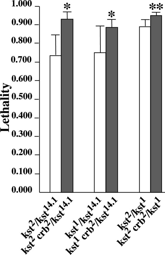
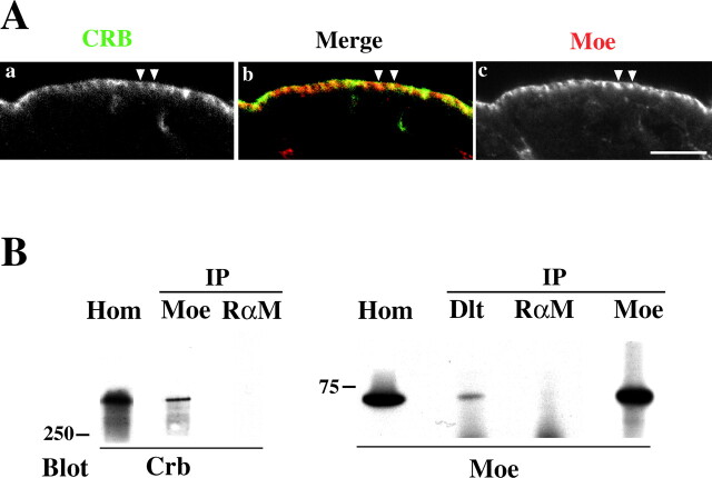
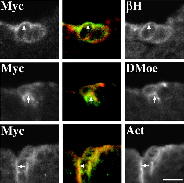
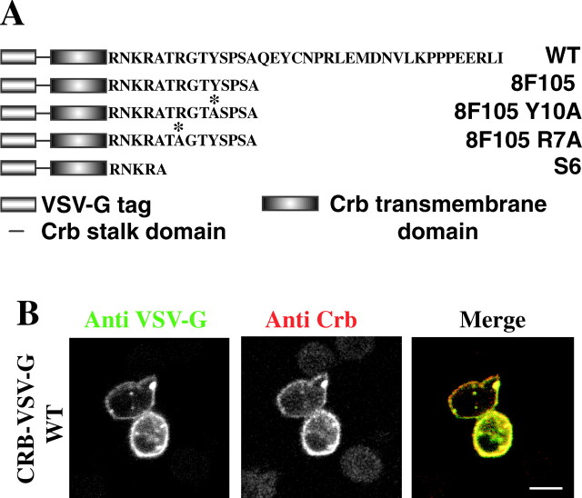
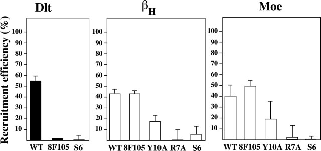
The apical transmembrane protein Crumbs is necessary for both cell polarization and the assembly of the zonula adherens (ZA) in Drosophila epithelia. The apical spectrin-based membrane skeleton (SBMS) is a protein network that is essential for epithelial morphogenesis and ZA integrity, and exhibits close colocalization with Crumbs and the ZA in fly epithelia. These observations suggest that Crumbs may stabilize the ZA by recruiting the SBMS to the junctional region. Consistent with this hypothesis, we report that Crumbs is necessary for the organization of the apical SBMS in embryos and Schneider 2 cells, whereas the localization of Crumbs is not affected in karst mutants that eliminate the apical SBMS. Our data indicate that it is specifically the 4.1 protein/ezrin/radixin/moesin (FERM) domain binding consensus, and in particular, an arginine at position 7 in the cytoplasmic tail of Crumbs that is essential to efficiently recruit both the apical SBMS and the FERM domain protein, DMoesin. Crumbs, Discs lost, betaHeavy-spectrin, and DMoesin are all coimmunoprecipitated from embryos, confirming the existence of a multimolecular complex. We propose that Crumbs stabilizes the apical SBMS via DMoesin and actin, leading to reinforcement of the ZA and effectively coupling epithelial morphogenesis and cell polarity.


Figures

Figure 1.
Figure 1.
βH is mislocalized in crumbs8F105 embryos. (A) Schematic figure showing the organization of cell–cell junctions in the ectoderm of Drosophila. Arm, armadillo; SJ, septate junction; ZA, zonula adherens. Some key markers for the different structures are indicated. (B) Confocal micrographs showing part of the ectoderm from stage 11/12 embryos stained for βH (bottom) and Crumbs (top). In WT embryos, βH and Crumbs are highly concentrated in the apicolateral region of each cell (arrows). In crumbs8F105 embryos (crb8F105), βH is mislocalized to the whole apical membrane (asterisks), and to a lesser extent to the cytoplasm. Bar, 5 μm.
Figure 2.
Figure 2.
crumbs dominantly enhances karst lethality. Lethality is expressed as the fraction of Mendelian expectation. The values plotted were all estimated from multiple crosses with large sample sizes. From left to right the number of crosses/total number of flies scored were: 4/2,059; 19/10,013; 4/2,204; 5/3,655; 9/4,102; and 7/3,329. The presence of one mutant crumbs11A22 allele (crb2) significantly increased lethality in all genotypic combinations (* = P < 0.05; ** = P < 0.01). See Materials and methods for details on the statistical analysis of these data. Error bars represent 95% confidence intervals.
Figure 3.
Figure 3.
Coimmunoprecipitation experiments reveal a CRB–βH complex. WT embryo lysates were immunoprecipitated (IP) with antibodies against βH or Dlt or with rabbit anti–mouse antibodies (RαM) and probed on immunoblots (Blot) with antibodies against α-spectrin (α-Sp) or Crumbs (Crb). Crumbs and α-spectrin coprecipitate with βH. Migration of markers is indicated in kD. Hom, a whole embryo extract loaded on the same gel as a control.
Figure 4.
Figure 4.
DMoesin interacts with Crumbs and βH. (A) Confocal micrographs showing part of the ectoderm from stage 11/12 embryos stained for Crumbs (a) and DMoesin (c). DMoesin and Crumbs are concentrated in the apicolateral domain (arrowheads). (B) Crumbs coimmunoprecipitates with DMoesin. Protein extracts from wild type embryos were immunoprecipitated (IP) with antibodies against DMoesin (Moe), Dlt, or rabbit anti–mouse antibodies (RαM). The resulting immunoprecipitates were immunoblotted with antibodies against DMoesin (Moe) or Crumbs (Crb). Migration of markers is indicated in kD. Hom, whole embryo extract as control.
Figure 5.
Figure 5.
Overexpression of Myc-intraWTleads to the redistribution of βH, DMoesin, and actin. Confocal micrographs of part of the epidermis of a stage 13/14 embryo. Cells at the posterior margin of each segment are expressing Myc-intraWT driven by an engrailed-Gal4 driver. Embryos were stained for Myc and βH, Dmoesin, (DMoe), or actin (Act) as indicated. βH, DMoesin, and actin are all redistributed along with Myc-intraWT (arrows). Bar, 5 μm.
Figure 6.
Figure 6.
Expression of CRB–VSV-G WT in S2 cells. (A) Sequences of the CRB–VSV-G fusion proteins expressed in this study. CRB–VSV-G WT is a fusion of the VSV-G epitope with the stalk region, transmembrane domain and the intracellular domain of Crumbs. Stop mutations in position 6 or 15 truncate the intracellular domain of Crumbs resulting in a cytoplasmic domain of 5 and 14 amino acids in variants CRB–VSV-G S6 and CRB–VSV-G 8F105, respectively. CRB–VSV-G 8F105 Y10A and R7A are CRB–VSV-G 8F105 constructs with point mutations (asterisk) replacing Tyr10 and Arg7 with an alanine, respectively. (B) CRB–VSV-G WT expression was induced in stably transfected S2 cells and cells were fixed and double labeled with a mouse anti–VSV-G and a rabbit anticytoplasmic domain of Crumbs antibodies followed by FITC-conju- gated anti–mouse (left) and TRITC- conjugated anti–rabbit (middle) antibodies. The two antibodies stained the same subcellular structures and in particular the plasma membrane (right). Bar, 10 μm.
Figure 7.
Figure 7.
Dlt, βH, and DMoesin colocalize with capped CRB–VSV-G WT in transfected S2 cells. CRB–VSV-G WT (A–C, top) or S6 (A–C, bottom) were transiently expressed in S2 cells, followed by capping and staining with mouse anti–VSV-G antibody and fluorescein-conjugated anti–mouse antibody before fixation. After fixation and permeabilization, cells were additionally stained for either Dlt (A, Dlt), βH (B, βH), or DMoesin (C, Moe). Dlt, βH, and DMoesin were all redistributed to capped sites with CRB–VSV-G WT indicating a connection between these proteins and the cytoplasmic domain of Crumbs in S2 cells (arrows highlight specific examples). Bar, 10 μm.
Figure 8.
Figure 8.
Quantitative analysis of βH recruitment to CRB–VSV-G cap sites. S2 cells expressing the different CRB–VSV-G constructs were scored for Dlt, βH, or DMoesin (Moe) colocalization after capping with anti–VSV-G and secondary antibodies. Results are expressed as the mean percentage of cocapping seen for each protein from three independent experiments (except for Moe 8F105, Y10A, and S6, which were performed twice, and Dlt 8F105 which was done once).
Figure 9.
Figure 9.
Model for the protein interactions in the Crumbs complex. A model summarizing all the published data and those presented in this study is drawn to show the interactions occurring inside the Crumbs complex. The amino acids playing a crucial role for the interactions are indicated. See Discussion for details.

References

    1. Bachmann, A., M. Schneider, E. Theilenberg, F. Grawe, and E. Knust. 2001. Drosophila Stardust is a partner of Crumbs in the control of epithelial cell polarity. Nature. 414:638–643. - PubMed
    1. Bennett, V., and A.J. Baines. 2001. Spectrin and ankyrin-based pathways: metazoan inventions for integrating cells into tissues. Physiol. Rev. 81:1353–1392. - PubMed
    1. Bhat, M.A., S. Izaddoost, Y. Lu, K.O. Cho, K.W. Choi, and H.J. Bellen. 1999. Discs Lost, a novel multi-PDZ domain protein, establishes and maintains epithelial polarity. Cell. 96:833–845. - PubMed
    1. Bilder, D. 2001. Cell polarity: squaring the circle. Curr. Biol. 11:R132–R135. - PubMed
    1. Bilder, D., and N. Perrimon. 2000. Localization of apical epithelial determinants by the basolateral PDZ protein Scribble. Nature. 403:676–680. - PubMed

Publication types

MeSH terms