. 2002 Oct;22(19):6809-19.
doi: 10.1128/MCB.22.19.6809-6819.2002.

Mitotic phosphorylation of chromosomal protein HMGN1 inhibits nuclear import and promotes interaction with 14.3.3 proteins

Affiliations

Mitotic phosphorylation of chromosomal protein HMGN1 inhibits nuclear import and promotes interaction with 14.3.3 proteins

Marta Prymakowska-Bosak et al. Mol Cell Biol. 2002 Oct.

Abstract

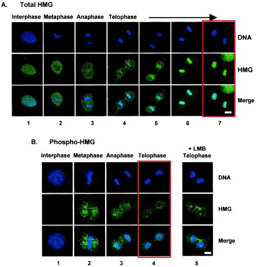
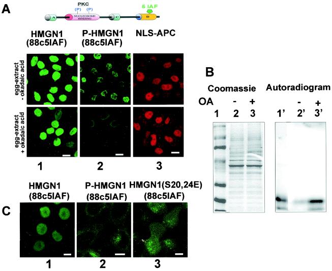
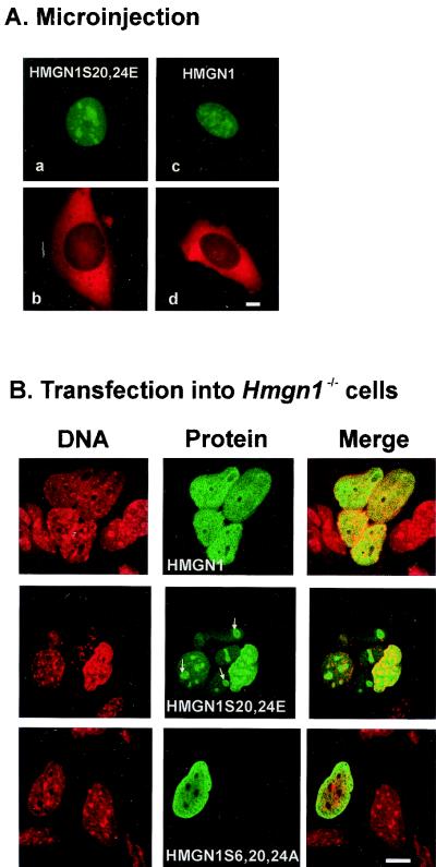
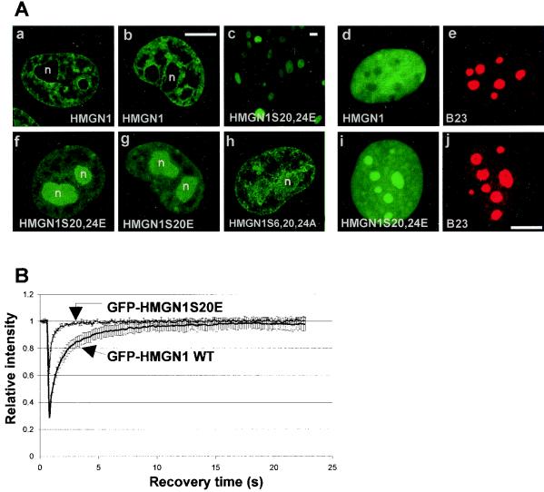
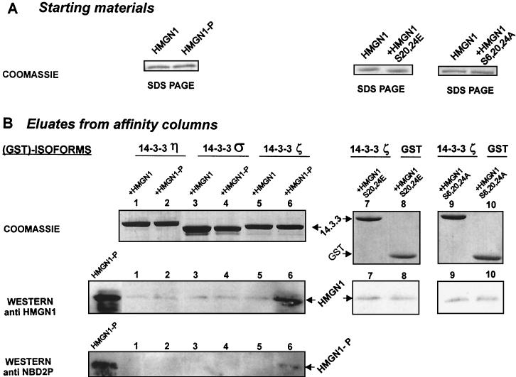
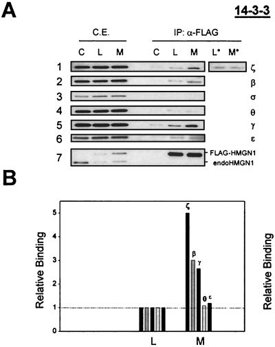
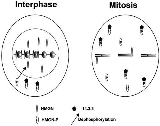
Progression through mitosis is associated with reversible phosphorylation of many nuclear proteins including that of the high-mobility group N (HMGN) nucleosomal binding protein family. Here we use immunofluorescence and in vitro nuclear import studies to demonstrate that mitotic phosphorylation of the nucleosomal binding domain (NBD) of the HMGN1 protein prevents its reentry into the newly formed nucleus in late telophase. By microinjecting wild-type and mutant proteins into the cytoplasm of HeLa cells and expressing these proteins in HmgN1(-/-) cells, we demonstrate that the inability to enter the nucleus is a consequence of phosphorylation and is not due to the presence of negative charges. Using affinity chromatography with recombinant proteins and nuclear extracts prepared from logarithmically growing or mitotically arrested cells, we demonstrate that phosphorylation of the NBD of HMGN1 promotes interaction with specific 14.3.3 isotypes. We conclude that mitotic phosphorylation of HMGN1 protein promotes interaction with 14.3.3 proteins and suggest that this interaction impedes the reentry of the proteins into the nucleus during telophase. Taken together with the results of previous studies, our results suggest a dual role for mitotic phosphorylation of HMGN1: abolishment of chromatin binding and inhibition of nuclear import.


Figures

FIG. 1.
FIG. 1.
Relocalization of HMGN1, but not of phosphorylated HMGN1, with nuclear DNA during telophase. Confocal immunofluorescence microscopy of mouse embryonic fibroblasts with antibodies elicited against either the C-terminal domain of mouse HMGN1 (A) or against the phosphorylated NBD of HMGN1 (B). The locations of the antibodies were determined by indirect immunofluorescence with fluorescein-labeled goat anti-rabbit immunoglobulin G. Note the increased colocalization of HMGN1 with DNA and the gradual decrease in extrachromosomal HMGN1 as the cell progresses through telophase. The panels boxed in red depict cells in late telophase. Note the differences in color of the nucleus in the merged panels in panels A and B. In panel B, panels 5 are confocal images from cells grown in the presence of leptomycin B (LMB). Bars, 10 μm.
FIG. 2.
FIG. 2.
Phosphorylation of HMGN blocks nuclear import. (A) Inhibition of phosphatase activity blocks nuclear import of phosphorylated HMGN1. Fluorescein-labeled HMGN1 is diagrammed at the top of panel A; 5IAF is the fluorescent acetamide-fluorescein attached to the C-terminal domain of the protein, P denotes PKC-phosphorylated serines in the NBD of HMGN1, and NLC-APC is the NLS-allophycocyanin conjugate. Confocal images visualizing the import of fluorescein-labeled HMGN1 into permeabilized HEp-2 cells, mediated by a X. laevis egg extract that contained or did not contain okadaic acid. In the absence of okadaic acid, the phosphorylated HMGN1 protein (P-HMGN1) was imported into the nucleus less efficiently than the nonphosphorylated HMGN1, as evidenced by the intensity of the fluorescent signal (compare panels 1 and 2 in top row). In the presence of okadaic acid, the import of the phosphorylated protein but not that of the nonphosphorylated protein was totally blocked (compare panels 1 and 2 in bottom row). (B) Stability of HMGN1 phosphorylation in Xenopus egg extracts treated with okadaic acid and analyzed by SDS-PAGE. 5IAF-labeled HMGN1, phosphorylated with 32P ATP by PKC, was incubated in Xenopus egg extract that contained (+) or did not contain (−) okadaic acid (OA). An aliquot of the phosphorylated protein was added to the molecular size markers (lane 1). Note that okadaic acid inhibited the phosphatase activity in the egg extract (compare lane 2′ to lane 3′). (C) Negative charges in the NBD do not inhibit nuclear import. Import of fluorescently labeled HMGN1, phosphorylated HMGN1 (P-HMGN1), or the negatively charged HMGN1S20,24E mutant with extracts containing okadaic acid. Note that the phosphorylated HMGN1 protein does not enter the nucleus, but the double point mutant does (compare panel 2 to panel 3). Bars, 20 μm.
FIG. 3.
FIG. 3.
Negative charges in the HMGN NBD do not inhibit nuclear import. (A) Fluorescence microscopy of mouse embryonic fibroblasts injected with solutions containing fluorescently labeled (green) wild-type or mutant HMGN1 and high-molecular-weight dextran labeled with Texas red. The dextran remains in the cytoplasm, while both proteins enter the nucleus. (B) Confocal laser scanning fluorescence microscopy visualizing the location of either the DNA or the transfected proteins in HmgN1−/− mouse fibroblasts. The proteins expressed in the various cells are indicated in the figure. Since the endogenous protein is not expressed in these cells, the transfected protein was visualized by indirect immunofluorescence with antibodies specific to HMGN1. Note that the wild-type protein is evenly distributed throughout the interphase nucleus, while the double mutant with a negatively charged NBD forms aggregates (white arrows). Bars, 20 μm.
FIG. 4.
FIG. 4.
Mislocalization of negatively charged HMGN1 mutants. Wild-type HMGN1 and GFP-labeled HMGN1 mutant proteins expressed in HeLa cells are shown. The type of GFP-labeled protein is indicated in the various panels. Note that wild-type HMGN1 is distributed throughout the entire nonnucleolar space in the nucleus (a and b). The negatively charged HMGN1S20,24E mutant is efficiently expressed in and enters the nucleus in all the HeLa cells (c and f). This double point mutant mislocalizes to the nucleolus (f), as does the single point mutant (g). The triple mutant HMGN1S6,20,24A, which is not negatively charged, is distributed throughout the entire nucleus (h). The nucleolar localization in living cells was independently verified by immunofluorescence with anti-B23 using fixed cells (cells expressing wild-type protein [d and e] and cells expressing mutant HMGN1 [i and j]). n, nucleolus. Bars, 20 μm (b and j) and 50 μm (c). (B) FRAP analysis of the intranuclear mobility of the wild-type (WT) HMGN1 and single point mutant HMGN1S20E. Note that the fluorescence recovery of the mutant is faster than that of the wild type, indicating that the mutant is more mobile.
FIG. 5.
FIG. 5.
Specific binding of phosphorylated HMGN1 (HMGN1-P) to 14.3.3ζ. (A) Polyacrylamide gel analysis of the starting materials, applied to the affinity columns. The SDS-polyacrylamide gels demonstrate that the solutions contained equal amounts of protein. (B) Analysis of the eluates from the 14.3.3 affinity columns. Either nonphosphorylated or PKC-phosphorylated HMGN1 was added to the GST-14.3.3 isoforms indicated. Coomassie blue staining of SDS-polyacrylamide gels of the eluates indicates that each fraction contained equal amounts of 14.3.3 protein. The results of Western analysis indicate a specific interaction between phosphorylated HMGN1 (HMGN1-P) and 14.3.3ζ. Equal trace amounts of HMGN1S20,24E bound to agarose beads containing 14.3.3ζ or GST (lanes 7 and 8). The HMGN1S6,20,24A mutant also does not bind to 14.3.3ζ (lanes 9 and 10).
FIG. 6.
FIG. 6.
Coimmunoprecipitation reveals preferential associations of HMGN1 with specific isoforms of 14.3.3 proteins in mitotic cells. (A) Total cell extracts (C.E.) prepared from control cells (C), from logarithmically growing cells expressing FLAG-tagged HMGN1 (L), or from mitotic cells expressing the FLAG-tagged HMGN1 (M) were treated with immobilized anti-FLAG antibodies (α-FLAG). Lanes L* and M* contain extracts not treated with phosphatase inhibitors and the mitotic HMGNs are not phosphorylated. The proteins in the extracts (C.E.) and the materials bound to the affinity column (IP: α-FLAG) were fractionated by SDS-PAGE, and the contents of the various 14.3.3 isoforms were visualized by immunoblotting with specific antibodies. The antibodies used to develop the blots are indicated on the right under the 14.3.3 heading. Each of the three C.E. lanes, lanes C, L, and M, contained 5 μg of cell extract. In the IP: α-FLAG lanes, each lane contained 1/40th of the material recovered from the affinity columns. Each strip is a separate gel. The lowest strip shows a Western blot with anti-HMGN1 antibody, demonstrating the relative levels of HMGN1 and FLAG-HMGN1 in the cells. IP, immunoprecipitation. (B) Quantification of the relative enrichment of the signal obtained with the various anti-14.3.3 antibodies. In each case, the signal obtained with the FLAG-bound proteins from the logarithmically growing cells (L) was set at 1.0 (dotted line). In the mitotic cell preparation (M), about 65% of the cells were in metaphase.
FIG. 7.
FIG. 7.
Model of the effect of mitotic phosphorylation on the chromatin interaction and intracellular trafficking of HMGN proteins. In the interphase nucleus, nonphosphorylated proteins are found either associated with nucleosomes or in the nucleoplasm. In mitotic cells, the NBDs of most of the HMGN proteins are phosphorylated. The phosphorylated protein is not bound to chromosomes and can form a complex with 14.3.3. At the end of mitosis, the phosphorylated protein is temporarily sequestered in the cytoplasm by its association with 14.3.3 protein. Nuclear entry is associated with dephosphorylation of the NBD. HMGN-P, phosphorylated HMGN.

References

    1. Bakiri, L., D. Lallemand, E. Bossy-Wetzel, and M. Yaniv. 2000. Cell cycle-dependent variations in c-Jun and JunB phosphorylation: a role in the control of cyclin D1 expression. EMBO J. 19:2056-2068. - PMC - PubMed
    1. Boulikas, T. 1995. Phosphorylation of transcription factors and control of the cell cycle. Crit. Rev. Eukaryot. Gene Expr. 5:1-77. - PubMed
    1. Brunet, A., F. Kanai, J. Stehn, J. Xu, D. Sarbassova, J. V. Frangioni, S. N. Dalal, J. A. DeCaprio, M. E. Greenberg, and M. B. Yaffe. 2002. 14-3-3 transits to the nucleus and participates in dynamic nucleocytoplasmic transport. J. Cell Biol. 156:817-828. - PMC - PubMed
    1. Bustin, M. 2001. Chromatin unfolding and activation by HMGN(*) chromosomal proteins. Trends Biochem. Sci. 26:431-437. - PubMed
    1. Bustin, M. 1999. Regulation of DNA-dependent activities by the functional motifs of the high-mobility-group chromosomal proteins. Mol. Cell. Biol. 19:5237-5246. - PMC - PubMed

Publication types

MeSH terms

LinkOut - more resources