. 2002 Oct;22(19):6820-30.
doi: 10.1128/MCB.22.19.6820-6830.2002.

Gene structure and functional analysis of the mouse nidogen-2 gene: nidogen-2 is not essential for basement membrane formation in mice

Affiliations

Gene structure and functional analysis of the mouse nidogen-2 gene: nidogen-2 is not essential for basement membrane formation in mice

Jürgen Schymeinsky et al. Mol Cell Biol. 2002 Oct.

Abstract

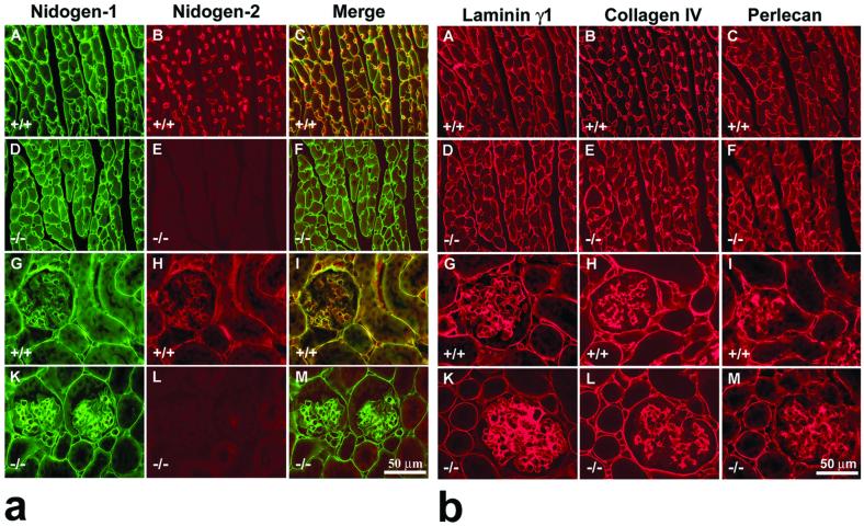
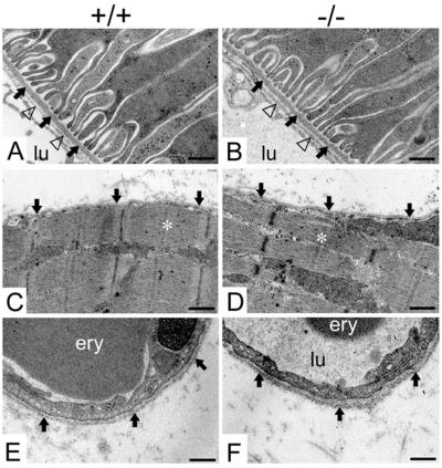
Nidogens are highly conserved proteins in vertebrates and invertebrates and are found in almost all basement membranes. According to the classical hypothesis of basement membrane organization, nidogens connect the laminin and collagen IV networks, so stabilizing the basement membrane, and integrate other proteins. In mammals two nidogen proteins, nidogen-1 and nidogen-2, have been discovered. Nidogen-2 is typically enriched in endothelial basement membranes, whereas nidogen-1 shows broader localization in most basement membranes. Surprisingly, analysis of nidogen-1 gene knockout mice presented evidence that nidogen-1 is not essential for basement membrane formation and may be compensated for by nidogen-2. In order to assess the structure and in vivo function of the nidogen-2 gene in mice, we cloned the gene and determined its structure and chromosomal location. Next we analyzed mice carrying an insertional mutation in the nidogen-2 gene that was generated by the secretory gene trap approach. Our molecular and biochemical characterization identified the mutation as a phenotypic null allele. Nidogen-2-deficient mice show no overt abnormalities and are fertile, and basement membranes appear normal by ultrastructural analysis and immunostaining. Nidogen-2 deficiency does not lead to hemorrhages in mice as one may have expected. Our results show that nidogen-2 is not essential for basement membrane formation or maintenance.


Figures

FIG. 1.
FIG. 1.
Domain structure (A) and gene locus (B) of murine nidogen-2, and characterization of the gene trap vector insertion site in the mutant nidogen-2 allele (C). (A) Schematic drawing of the domain structure of nidogen-2. Lines define the exons encoding the predicted globular domains G1, G2, and G3 and the connecting elements, the flexible link and the rigid rod. Numbers refer to amino acid positions of corresponding domain borders. (B) The organization of the murine nidogen-2 gene is shown. The filled boxes representing the 21 translated exons are drawn to scale. The exon numbers are indicated on the top of the boxes. The translational start and stop codon (ATG and TGA) and the poly(A) adenylation signal AATAAA are indicated. The lines at bottom represent the inserts of overlapping λ phages and genomic PCR clone pEX17/18 carrying the complete nidogen-2 gene. Only the EcoRI restriction sites are indicated on the phage maps. (C) The schematic drawings represent the wild-type and trapped alleles of nidogen-2. The vertical arrow indicates the insertion site of the gene trapping vector pGT1.8TM within intron 4. The positions of the probes (probes A, B, and lacZ) used in genomic Southern blot analysis are indicated as boxes. The positions of the primers used in PCR assays are depicted by horizontal arrowheads. Key DNA restriction fragments (wild-type, 1.5-kb PstI fragment; mutant, 3.5-kb PstI fragment) detected by Southern blot and DNA fragments (wild-type, 1.1 kb; mutant, 0.7 kb) generated by PCR assays for genotyping mice are shown as horizontal bars. Abbreviations: E, EcoRI; H, HindIII; K, KpnI; P, PstI; S, SalI; X, XbaI; βgeo, β-galactosidase-neomycin fusion gene; en-2, engrailed 2 gene; SA, splice acceptor site; SD, splice donor site; TM, transmembrane domain of CD4.
FIG. 2.
FIG. 2.
Characterization of the gene trap vector insertion site by Southern blotting (A) and genotype analyses by Southern blotting (B) and PCR (C). (A) Genomic DNA digested with different restriction endonucleases was analyzed by hybridization with probes A and B. The probes detect in genomic DNA derived from wild-type (+/+) mice one single band, whereas in genomic DNA derived from heterozygous mutant (+/−) in most cases two specific bands corresponding to both alleles are readily recognized. (B) Southern blots of genomic DNA digested with the restriction endonuclease PstI were analyzed by hybridization with probe B. The 1.5-kb band derived from the wild-type allele and the 3.5-kb band specific for the mutant allele are indicated on the right. Genotypes with the expected specific hybridization patterns of wild-type (+/+), heterozygous (+/−) and homozygous (−/−) mutant mice are shown. (C) PCR analysis of genomic DNA as template and with primers P1, ND70, and ND79. The 1.1-kb band, the product of primers P1 and ND70, represents the wild-type allele (+/+); the 0.7-kb band, the product of primers ND79 and ND70, represents the mutant allele (−/−). The markers on the left represent a 1-kb ladder (GIBCO-BRL). Abbreviations: E, EcoRI; H, HindIII; K, KpnI; P, PstI; S, SalI; X, XbaI.
FIG. 3.
FIG. 3.
Northern blot analysis of RNA isolated from heart, lung, kidney, and skeletal muscle from wild-type and mutant (−/−) nidogen-2 mice. (A) Poly(A)+ RNA fractionated by agarose gel electrophoresis (4 μg/lane) and transferred to nylon membranes was hybridized to nidogen-2-specific probe C. (B) After stripping of the radioactivity, the same blot was rehybridized with a nidogen-1-specific probe and a GAPDH-specific probe (not shown). (C) In a parallel blot hybridization was performed using a lacZ-specific probe. The markers on the left represent an RNA ladder (GIBCO-BRL). Abbreviations: H, heart; K, kidney; L, lung; M, skeletal muscle.
FIG. 4.
FIG. 4.
Western blot analysis of protein extracts of heart, lung and skeletal muscle from normal (+/+) and mutant (−/−) mice. Extracted protein (60 μg) was fractionated by sodium dodecyl sulfate-polyacrylamide gel electrophoresis, transferred to Immobilon membranes, and stained for nidogens with antiserum to nidogen-1 (A) and antiserum to nidogen-2 (B). (A) Both wild-type and nidogen-2 (−/−) extracts contain nidogen-1-specific bands of 150, 130, and 100 kDa (closed arrowheads) in apparently normal and comparable amounts. (B) The nidogen-2 (−/−) mice lack the nidogen-2-specific bands (open arrowheads). The gels were calibrated by globular proteins, denoted in the margins. Abbreviations: H, heart; L, lung; M, skeletal muscle.
FIG. 5.
FIG. 5.
Indirect immunofluorescence microscopy of cardiac muscle (A to F) and kidney (G to M) from adult control (+/+) and nidogen-2 (−/−) mice. (a) Double-staining immunofluorescence was applied to sections of cardiac muscle and kidney using rat monoclonal antibodies to mouse nidogen-1 (A, D, G, and K) (green signal) and affinity-purified antibodies to nidogen-2 (B, E, H, and L) (red signal). (C, F, I, and M) Both color channels were merged to demonstrate codistribution (yellow signal) of both immunofluorescence staining signals in the corresponding section. (A and D) Prominent nidogen-1-specific staining is shown around cardiomyocytes and capillaries in both wild-type (A) and mutant (D) sections. (B) In control sections, staining for nidogen-2 was typically prominent around capillaries, whereas the staining intensities around cardiomyocytes are much weaker. This aspect is well demonstrated by showing the merged images (C). (E) In the tissue section of the mutant heart, no nidogen-2-specific staining was observed. (G to I) In the sections of wild-type kidney, a close colocalization of both nidogens was mainly demonstrated in the BM zones of the proximal and distal tubuli, glomeruli, the Bowman's capsule, and blood vessels. In the tissue section of the mutant kidney (K), nidogen-1 staining is similar to nidogen-1 immunofluorescence in the wild-type section (G), whereas no nidogen-2-specific staining was observed in panel L. Bar, 50 μm. (b) Single immunofluorescence microscopy of cardiac muscle (A to F) and kidney (G to M) from adult control (+/+) and nidogen-2 (−/−) mice was applied on consecutive sections using rabbit antisera as primary antibodies recognizing BM proteins laminin γ1 (A, D, G, and K), collagen IV (B, E, H, and L), or perlecan (C, F, I, and M). Cy3-conjugated secondary antibodies were used to visualize specific stainings. Prominent staining for all three BM proteins is shown in wild-type and mutant sections. Immunofluorescence staining intensities in control and mutant tissues appear to be maintained at equal levels. Bar, 50 μm.
FIG. 6.
FIG. 6.
Electron micrographs of ultrathin sections of the kidney cortex (A and B), the hind limb soleus muscle (C and D), and capillaries (E and F) from adult wild-type (A, C, and E) and homozygous mutant (B, D, and F) animals. (A and B) The BM of a proximal tubule is marked by arrows, and the endothelial BM is indicated by triangles. (C and D) The BM of the myocyte is marked by arrows, and a sarcomere is marked by a white asterisk. (E and F) Arrows point to the BM of a capillary of the soleus muscle. Abbreviations: ery, erythrocyte; lu, capillary lumen. Bars, 0.25 μm (A to D) and 0.2 μm (E and F).

Similar articles

Cited by

References

    1. Arikawa-Hirasawa, E., H. Watanabe, H. Takami, J. R. Hassell, and Y. Yamada. 1999. Perlecan is essential for cartilage and cephalic development. Nat. Genet. 23:354-358. - PubMed
    1. Bork, P., A. K. Downing, B. Kieffer, and I. D. Campbell. 1996. Structure and distribution of modules in extracellular proteins. Q. Rev. Biophys. 29:119-167. - PubMed
    1. Brennan, J., and W. C. Skarnes. 1999. Gene trapping in mouse embryonic stem cells. Methods Mol. Biol. 97:123-138. - PubMed
    1. Brown, J. C., T. Sasaki, W. Göhring, Y. Yamada, and R. Timpl. 1997. The C-terminal domain V of perlecan promotes β1 integrin-mediated cell adhesion, binds heparin, nidogen and fibulin-2 and can be modified by glycosaminoglycans. Eur. J. Biochem. 250:39-46. - PubMed
    1. Costell, M., E. Gustafsson, A. Aszodi, M. Mörgelin, W. Bloch, E. Hunziker, K. Addicks, R. Timpl, and R. Fässler. 1999. Perlecan maintains the integrity of cartilage and some basement membranes. J. Cell Biol. 147:1109-1122. - PMC - PubMed

Publication types

MeSH terms

LinkOut - more resources