. 2002 Oct;184(19):5282-92.
doi: 10.1128/JB.184.19.5282-5292.2002.

A new modified ortho cleavage pathway of 3-chlorocatechol degradation by Rhodococcus opacus 1CP: genetic and biochemical evidence

Affiliations

A new modified ortho cleavage pathway of 3-chlorocatechol degradation by Rhodococcus opacus 1CP: genetic and biochemical evidence

Olga V Moiseeva et al. J Bacteriol. 2002 Oct.

Abstract

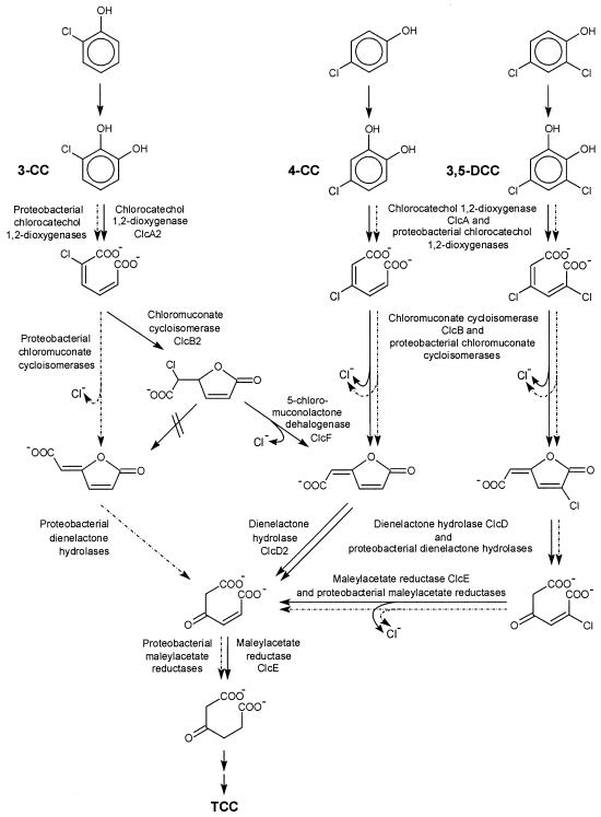
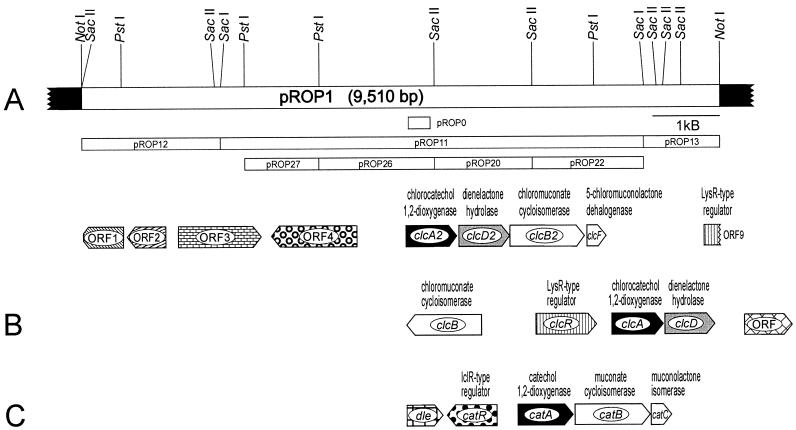
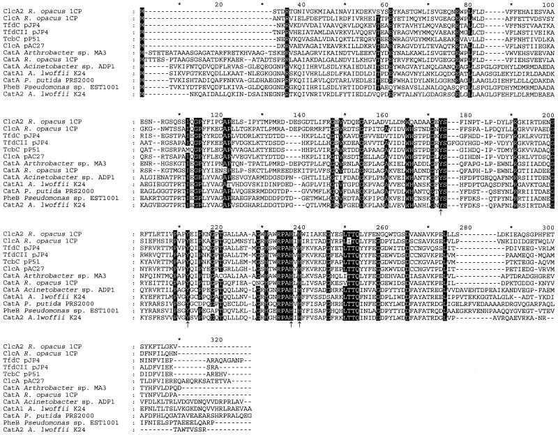
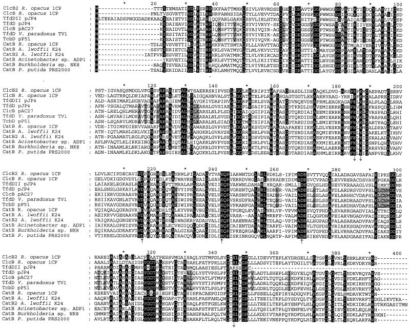
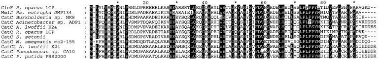
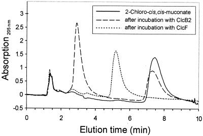
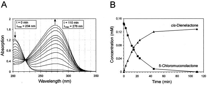
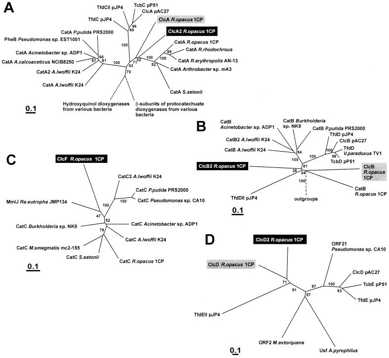
The 4-chloro- and 2,4-dichlorophenol-degrading strain Rhodococcus opacus 1CP has previously been shown to acquire, during prolonged adaptation, the ability to mineralize 2-chlorophenol. In addition, homogeneous chlorocatechol 1,2-dioxygenase from 2-chlorophenol-grown biomass has shown relatively high activity towards 3-chlorocatechol. Based on sequences of the N terminus and tryptic peptides of this enzyme, degenerate PCR primers were now designed and used for cloning of the respective gene from genomic DNA of strain 1CP. A 9.5-kb fragment containing nine open reading frames was obtained on pROP1. Besides other genes, a gene cluster consisting of four chlorocatechol catabolic genes was identified. As judged by sequence similarity and correspondence of predicted N termini with those of purified enzymes, the open reading frames correspond to genes for a second chlorocatechol 1,2-dioxygenase (ClcA2), a second chloromuconate cycloisomerase (ClcB2), a second dienelactone hydrolase (ClcD2), and a muconolactone isomerase-related enzyme (ClcF). All enzymes of this new cluster are only distantly related to the known chlorocatechol enzymes and appear to represent new evolutionary lines of these activities. UV overlay spectra as well as high-pressure liquid chromatography analyses confirmed that 2-chloro-cis,cis-muconate is transformed by ClcB2 to 5-chloromuconolactone, which during turnover by ClcF gives cis-dienelactone as the sole product. cis-Dienelactone was further hydrolyzed by ClcD2 to maleylacetate. ClcF, despite its sequence similarity to muconolactone isomerases, no longer showed muconolactone-isomerizing activity and thus represents an enzyme dedicated to its new function as a 5-chloromuconolactone dehalogenase. Thus, during 3-chlorocatechol degradation by R. opacus 1CP, dechlorination is catalyzed by a muconolactone isomerase-related enzyme rather than by a specialized chloromuconate cycloisomerase.


Figures

FIG. 1.
FIG. 1.
Degradative pathways for 3-chlorocatechol (3-CC), 4-chlorocatechol (4-CC), and 3,5-dichlorocatechol (3,5-DCC), the central intermediates in chlorophenol degradation, as found in R. opacus 1CP (solid lines). Dashed arrows show the known proteobacterial 3-chlorocatechol pathway for comparison. Enzyme names and their designations as gene products are given.
FIG. 2.
FIG. 2.
(A) Restriction map of the insert of pROP1 carrying 9,510 bp of R. opacus 1CP DNA (white bar) in the multiple cloning site of pBluescript II SK(+) (black bars). Shown below the map are the most important subclones and the probe pROP0. The locations and orientations of ORFs on pROP1 are indicated. (B and C) For comparison, R. opacus 1CP gene clusters for 4-chloro- and 3,5-dichlorocatechol catabolism (B) and for catechol catabolism (C) are shown. Homologous genes are presented in the same shade.
FIG. 3.
FIG. 3.
Sequence alignment of chlorocatechol 1,2-dioxygenases (ClcA, TfdC, and TcbC) and catechol 1,2-dioxygenases (CatA and PheB). The alignment was calculated using all sequences in Fig. 8 by using ClustalX1.8. Sequences not shown here were removed afterwards, and resulting column gaps were eliminated. Numbers above the sequences refer to positions in the alignment, not in individual sequences. Amino acids in positions in which at least 11 of the 13 sequences are identical are highlighted. Amino acids involved in iron binding are indicated by arrows (46). Amino acids shaded in gray are exclusively conserved in proteobacterial chloromuconate cycloisomerases able to dechlorinate 2-chloro-cis,cis-muconate. Accession numbers for the published sequences are as follows: ClcA from R. opacus 1CP, AF003948; TfdC from pJP4, M35097; TfdCII from pJP4, U16782; TcbD from pP51, M57629; ClcA from pAC27, M16964; CatA from Arthrobacter sp. strain mA3, AJ000187; CatA from R. opacus 1CP, X99622; CatA from Acinetobacter sp. strain ADP1, AF009224; CatA from A. lwoffii K24, U77658; CatA from P. putida PRS2000, U12557; PheB from Pseudomonas sp. strain EST1001, M57500; and CatA2 from Acinetobacter lwoffii K24, U77659.
FIG. 4.
FIG. 4.
Alignment of chloromuconate cycloisomerases (ClcB, TfdD, and TcbD) and muconate cycloisomerases (CatB). The alignment was calculated by using ClustalX1.8 with the outgroups shown in Fig 8. Outgroups and column gaps resulting from outgroup removal are not shown. Numbers above the sequences refer to positions in the alignment, not in individual sequences. Residues involved in manganese coordination (upward-pointing arrows) and residues directly involved in the enzyme mechanism (downward-pointing arrows) are indicated (10, 12, 14). Amino acids in positions in which at least 11 of the 13 sequences are identical are highlighted. Accession numbers for the published sequences are as follows: ClcB from R. opacus 1CP, AF003948; TfdDII from pJP4, U16782; TfdD from pJP4, M35097; ClcB from pAC27, M16964 (corrected as published by Vollmer et al. [48]); TfdD from Variovorax paradoxus TV1, AB028643; TcbD from pP51, M57629; CatB from R. opacus 1CP, X99622; CatB from A. lwoffii K24, U77658; CatB2 from A. lwoffii K24, U77659; CatB from Acinetobacter sp. strain ADP1, AF009224; CatB from Burkholderia sp. strain NK8, AB024746; and CatB from P. putida PRS2000, U12557.
FIG. 5.
FIG. 5.
Sequence alignment of 5-chloromuconolactone dehalogenase (ClcF) with muconolactone isomerases as calculated by using ClustalX1.8 and used for dendrogram calculations (Fig. 8). Numbers above the sequences refer to positions in the alignment, not in individual sequences. Amino acids in positions in which at least 10 of the 11 sequences are identical are highlighted. Accession numbers for the published sequences are as follows: MmlJ (methylmuconolactone isomerase) from R. eutropha JMP134, X99639; CatC from Burkholderia sp. strain NK8, AB024746; CatC from Acinetobacter sp. strain ADP1, AF009224; CatC from A. lwoffii K24, U77658; CatC from R. opacus 1CP, X99622; CatC from Streptomyces setonii, AF277051; CatC from Mycobacterium smegmatis mc2-155, AF144091; CatC2 from A. lwoffii K24, U77659; CatC from P. putida PRS2000, U12557; and CatC from Pseudomonas sp. strain CA10, AB047272.
FIG. 6.
FIG. 6.
Overlaid HPLC chromatograms of 2-chloro-cis,cis-muconate before and after sequential incubation with chloromuconate cycloisomerase ClcB2 and with the 5-chloromuconolactone-converting enzyme ClcF. At least 0.1 U of the homogeneous enzymes was added in sequence to a 1-ml assay mixture containing 0.2 μmol of 2-chloro-cis,cis-muconate and 50 μmol of Tris-HCl buffer (pH 7.5). Samples, which were taken from this mixture before enzyme addition, 10 min after incubation with ClcB2 alone, and 10 min after further incubation with ClcF, were quenched by addition of 10% (vol/vol) ortho-phosphoric acid and subjected to reversed-phase HPLC. The retention times for the following compounds at a flow rate of 1 ml/min were as follows: 5-chloromuconolactone, 2.92 min; cis-dienelactone, 5.20 min; and 2-chloro-cis,cis-muconic acid, 7.47 min. The signal at 1.5 min refers to the injection peak.
FIG. 7.
FIG. 7.
(A) Spectral changes during the ClcF-catalyzed conversion of 5-chloromuconolactone to cis-dienelactone. (B) Stoichiometry of the reaction, estimated by reversed-phase HPLC. A 0.2-μmol amount of 5-chloromuconolactone was incubated with 0.01 μg of purified ClcF (7 × 10⁻³ U as measured with 5-chloromuconolactone) in phosphate buffer (pH 7.2) at room temperature. UV spectra were recorded at 0, 0.5, 3, 6, 9, 12, 15, 18, 21, 24, 45, and 110 min. From an identical second reaction mixture, samples were taken at similar intervals. After quenching by addition of 10% (vol/vol) ortho-phosphoric acid, those samples were subjected to HPLC.
FIG. 8.
FIG. 8.
Dendrograms showing the relatedness of catechol 1,2-dioxygenases (CatA and PheB) and chlorocatechol 1,2-dioxygenases (ClcA, TfdC, and TcbC) (A), muconate cycloisomerase (CatB) and chloromuconate cycloisomerases (ClcB, TfdD, and TcbD) (B), muconolactone isomerases (CatC and MmIJ) and 5-chloromuconolactone dehalogenase (ClcF) (C), and dienelactone hydrolases (ClcD, TfdE, and TcbE) (D). The dendrograms were calculated using programs (Protdist and Fitch) of the PHYLIP package (version 3.5c) based on sequence alignments calculated by ClustalX1.8. Bootstrap values were derived from Seqboot (PHYLIP) calculations with 100 replicates. For the cycloisomerases, the muconolactone isomerases, and the dienelactone hydrolases, the complete alignments were used for dendrogram calculation (Fig. 4 and 5). For the dioxygenases, only positions 112 to 259 in Fig. 3 were used. Sequences examined in this study are highlighted black; sequences already known for chlorocatechol degradation in R. opacus 1CP are highlighted gray. Broken lines indicate that branches were cut. Accession numbers for catechol 1,2-dioxygenases not shown in Fig. 3 are as follows: CatA from A. calcoaceticus NCIB8250, Z36909; CatA from R. rhodochrous, AF043741; CatA from R. erythropolis AN-13, D83237; and CatA from S. setonii, AF277051. Accession numbers for the dienelactone hydrolases are as follows: ClcD from R. opacus 1CP, AF003948; TfdEII from pJP4, U16782; ORF21 from Pseudomonas sp. strain CA10, AB047548; ClcD from pAC27, M16964; TcbE from pP51, M57629; and TfdE from pJP4, M35097. Accession numbers for sequences used as outgroups are as follows. For panel A, hydroxyquinol dioxygenases from Burkholderia cepacia AC1100 (U19883), Ralstonia pickettii DTP0602 (D86544), and Arthrobacter sp. strain BA-5-17 (AB016258) and β-subunits of protocatechuate 3,4-dioxygenases from Pseudomonas sp. strain HR199 (Y18527), P. putida WCS358 (AJ295623), P. putida NCIMB9869 (U96339), alphaproteobacterium Y3F (AF253466), Agrobacterium tumefaciens A348 (U32867), and P. marginata ATCC10248 (U33634) were used. For panel B, N-acylamino acid racemases from Amycolatopsis orientalis subsp. lurida (AJ292519), Amycolatopsis sp. strain TS-1-60 (D30738), Deinococcus radiodurans R1 (AE001867), and Thermoplasma acidophilum DSM1728 (AL445063); mandelate racemase from P. putida (J05293); mandelate racemase-like protein from Sulfolobus solfataricus (AE006902); starvation-sensing protein RspA from E. coli (D90799); and putative muconate cycloisomerase YkfB from Bacillus subtilis (AJ002571) were used. For panel D, ORF2 from Methylobacterium extorquens AM1 (U72662) and Usf from Aquifex pyrophilus Ko15a (U17575), both with unknown function, were used.

Similar articles

Cited by

References

    1. Blasco, R., R. M. Wittich, M. Mallavarapu, K. N. Timmis, and D. H. Pieper. 1995. From xenobiotic to antibiotic, formation of protoanemonin from 4-chlorocatechol by enzymes of the 3-oxoadipate pathway. J. Biol. Chem. 270:29229-29235. - PubMed
    1. Bradford, M. M. 1976. A rapid and sensitive method for the quantitation of microgram quantities of protein utilizing the principle of protein-dye binding. Anal. Biochem. 72:248-254. - PubMed
    1. Cole, S. T., R. Brosch, J. Parkhill, T. Garnier, C. Churcher, D. Harris, S. V. Gordon, K. Eiglmeier, S. Gas, C. E. Barry III, F. Tekaia, K. Badcock, D. Basham, D. Brown, T. Chillingworth, R. Connor, R. Davies, K. Devlin, T. Feltwell, S. Gentles, N. Hamlin, S. Holroyd, T. Hornsby, K. Jagels, A. Krogh, J. McLean, S. Moule, L. Murphy, S. Oliver, J. Osborne, M. A. Quail, M. A. Rajandream, J. Rogers, S. Rutter, K. Seeger, S. Skelton, S. Squares, R. Squares, J. E. Sulston, K. Taylor, S. Whitehead, and B. G. Barrell. 1998. Deciphering the biology of Mycobacterium tuberculosis from the complete genome sequence. Nature 393:537-544. - PubMed
    1. Dorn, E., M. Hellwig, W. Reineke, and H.-J. Knackmuss. 1974. Isolation and characterization of a 3-chlorobenzoate degrading pseudomonad. Arch. Microbiol. 99:61-70. - PubMed
    1. Dorn, E., and H.-J. Knackmuss. 1978. Chemical structure and biodegradability of halogenated aromatic compounds. Two catechol 1,2-dioxygenases from a 3-chlorobenzoate-grown pseudomonad. Biochem. J. 174:73-84. - PMC - PubMed

Publication types

MeSH terms

Associated data

LinkOut - more resources