. 2002 Oct;184(19):5393-401.
doi: 10.1128/JB.184.19.5393-5401.2002.

Forespore signaling is necessary for pro-sigmaK processing during Bacillus subtilis sporulation despite the loss of SpoIVFA upon translational arrest

Affiliations

Forespore signaling is necessary for pro-sigmaK processing during Bacillus subtilis sporulation despite the loss of SpoIVFA upon translational arrest

Lee Kroos et al. J Bacteriol. 2002 Oct.

Abstract

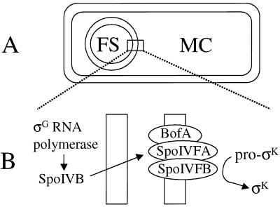
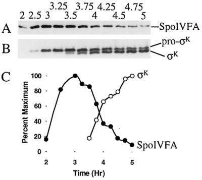
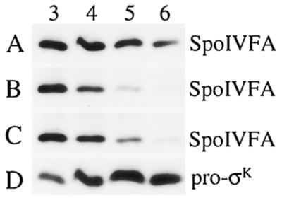
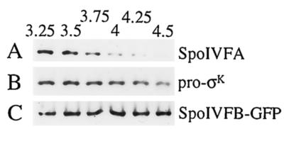
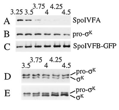
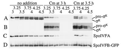
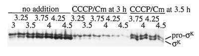
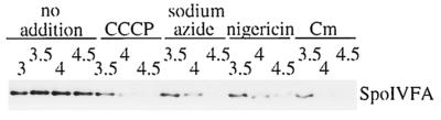
The sigmaK checkpoint coordinates gene expression in the mother cell with signaling from the forespore during Bacillus subtilis sporulation. The signaling pathway involves SpoIVB, a serine peptidase produced in the forespore, which is believed to cross the innermost membrane surrounding the forespore and activate a complex of proteins, including BofA, SpoIVFA, and SpoIVFB, located in the outermost membrane surrounding the forespore. Activation of the complex allows proteolytic processing of pro-sigmaK, and the resulting sigmaK RNA polymerase transcribes genes in the mother cell. To investigate activation of the pro-sigmaK processing complex, the level of SpoIVFA in extracts of sporulating cells was examined by Western blot analysis. The SpoIVFA level decreased when pro-sigmaK processing began during sporulation. In extracts of a spoIVB mutant defective in forespore signaling, the SpoIVFA level failed to decrease normally and no processing of pro-sigmaK was observed. Although these results are consistent with a model in which SpoIVFA inhibits processing until the SpoIVB-mediated signal is received from the forespore, we discovered that loss of SpoIVFA was insufficient to allow processing under certain conditions, including static incubation of the culture and continued shaking after the addition of inhibitors of oxidative phosphorylation or translation. Under these conditions, loss of SpoIVFA was independent of spoIVB. The inability to process pro-sigmaK under these conditions was not due to loss of SpoIVFB, the putative processing enzyme, or to a requirement for ongoing synthesis of pro-sigmaK. Rather, it was found that the requirements for shaking of the culture, for oxidative phosphorylation, and for translation could be bypassed by mutations that uncouple processing from dependence on forespore signaling. This suggests that ongoing translation is normally required for efficient pro-sigmaK processing because synthesis of the SpoIVB signal protein is needed to activate the processing complex. When translation is blocked, synthesis of SpoIVB ceases, and the processing complex remains inactive despite the loss of SpoIVFA. Taken together, the results suggest that SpoIVB signaling activates the processing complex by performing another function in addition to causing loss of SpoIVFA or by causing loss of SpoIVFA in a different way than when translation is blocked. The results also demonstrate that the processing machinery can function in the absence of translation or an electrochemical gradient across membranes.


Figures

FIG. 1.
FIG. 1.
σK checkpoint. (A) Diagram of a sporangium in which the forespore (FS) has been pinched off as a protoplast within the mother cell (MC). (B) Expanded view of the two membranes separating the forespore and mother cell, depicting BofA, SpoIVFA, and SpoIVFB in the outermost membrane surrounding the forespore. The three proteins form a complex in which SpoIVFB is inactive prior to signaling from the forespore (4, 5, 16, 30, 31, 33). σG RNA polymerase transcribes spoIVB in the forespore (3), and SpoIVB is secreted into the space between the two membranes (38), where it activates the processing complex, leading to the production of σK in the mother cell.
FIG. 2.
FIG. 2.
Levels of SpoIVFA, pro-σK, and σK during sporulation. (A) B. subtilis strain OR758 was induced to sporulate, samples were collected at the indicated times (hours) after sporulation was induced, and whole-cell extracts were subjected to Western blot analysis with antibodies against SpoIVFA. (B) The blot was stripped and reprobed with antibodies against pro-σK. (C) Quantification of the SpoIVFA (•) and σK (○) signal intensities from panels A and B, respectively.
FIG. 3.
FIG. 3.
Levels of SpoIVFA and pro-σK in mutants. B. subtilis strains were induced to sporulate, samples were collected at the indicated times (hours) after sporulation was induced, and whole-cell extracts were subjected to Western blot analysis with antibodies against SpoIVFA. (A) LK1 (spoIVBΔ::spc). (B) OR745 (spoIVFΔB::spc). (C) PY79 (spo+). (D) The blot in panel A was stripped and reprobed with antibodies against pro-σK.
FIG. 4.
FIG. 4.
Effect of discontinued shaking on pro-σK processing and on the levels of SpoIVFA and SpoIVFB-GFP. A portion of the OR758 (spoIVFB-gfp) culture used in the experiment shown in Fig. 2 was removed from shaking at T3, and incubation was continued at 37°C without shaking. (A) Samples were collected at the indicated times (hours) after sporulation was induced, and whole-cell extracts were subjected to Western blot analysis with antibodies against SpoIVFA. (B) The blot was stripped and reprobed with antibodies against pro-σK. (C) The blot was again stripped and this time reprobed with antibodies against GFP to detect the SpoIVFB-GFP fusion protein.
FIG. 5.
FIG. 5.
Effect of an uncoupler of oxidative phosphorylation on pro-σK processing and on the levels of SpoIVFA and SpoIVFB-GFP. A portion of the OR758 (spoIVFB-gfp) culture used in the experiment shown in Fig. 2 was removed at T3, CCCP (5 μM) was added, and shaking was continued. (A) Samples were collected at the indicated times (hours) after sporulation was induced, and whole-cell extracts were subjected to Western blot analysis with antibodies against SpoIVFA. (B) The blot was stripped and reprobed with antibodies against pro-σK. (C) The blot was again stripped and this time reprobed with antibodies against GFP to detect the SpoIVFB-GFP fusion protein. (D) In a separate experiment, the same strain used in the experiment shown in Fig. 2 was induced to sporulate, a portion of the culture was removed at T3.5, CCCP (5 μM) was added, and shaking was continued. Samples were collected at the indicated times (hours) after sporulation was induced, and whole-cell extracts were subjected to Western blot analysis with antibodies against pro-σK. (E) A portion of the culture used in the experiment shown in panel D was left untreated, and samples were analyzed as in panel D.
FIG. 6.
FIG. 6.
Effect of a protein synthesis inhibitor on pro-σK processing and on the levels of SpoIVFA and SpoIVFB-GFP. (A) B. subtilis strain OR758 was induced to sporulate, and the culture was split at T3 into portions to which nothing was added or Cm (200 μg/ml) was added immediately (Cm at 3 h) or after 30 min (Cm at 3.5 h). Shaking was continuous except when the culture was split and Cm was added. Samples were collected at the indicated times (hours) after sporulation was induced, and whole-cell extracts were subjected to Western blot analysis with antibodies against pro-σK. (B) Eightfold-longer exposure of part of the blot shown in panel A. (C) The blot was stripped and reprobed with antibodies against SpoIVFA. (D) The same samples used to produce the blot shown in panels A to C were used to produce another blot that was probed with antibodies against GFP to detect the SpoIVFB-GFP fusion protein.
FIG. 7.
FIG. 7.
Effect of a combination of inhibitors on pro-σK and σK levels. B. subtilis strain OR758 was induced to sporulate, and the culture was split at T3 into portions to which nothing was added or CCCP (5 μM) and Cm (200 μg/ml) were added immediately (CCCP/Cm at 3 h) or after 30 min (CCCP/Cm at 3.5 h). Shaking was continuous except when the culture was split and inhibitors were added. Samples were collected at the indicated times (hours) after sporulation was induced, and whole-cell extracts were subjected to Western blot analysis with antibodies against pro-σK.
FIG. 8.
FIG. 8.
Effect of inhibitors on the level of SpoIVFA in a spoIVB mutant. B. subtilis strain LK1 (spoIVBΔ::spc) was induced to sporulate, and the culture was split at T3 into portions to which nothing was added or CCCP (5 μM), sodium azide (0.2%), nigericin (1 μM), or Cm (200 μg/ml) was added. Shaking was continuous except when the culture was split and inhibitors were added. Samples were collected at the indicated times (hours) after sporulation was induced, and whole-cell extracts were subjected to Western blot analysis with antibodies against SpoIVFA.
FIG. 9.
FIG. 9.
Pulse-chase analysis of pro-σK processing. (A) The indicated B. subtilis strains were induced to sporulate, pulse-labeled with [35S]methionine at T3, and chased with a 1,000-fold molar excess of unlabeled methionine (lanes 1 to 3 and 7 to 12) or chased by collecting the cells by centrifugation and resuspending them in fresh medium with a 10,000-fold molar excess of unlabeled methionine (lanes 4 to 6). Numbers below the lanes indicate the length of the chase period in minutes. (B) The indicated strains were induced to sporulate, pulse-labeled with [35S]methionine at T3 (lanes 1 and 4), and chased with a 1,000-fold molar excess of unlabeled methionine for 30 min in the absence (lanes 2 and 5) or presence (lanes 3 and 6) of Cm (200 μg/ml). The numbers below the lanes indicate the σK/pro-σK ratio based on quantification of the [35S]methionine-labeled protein bands. (C) The indicated strains were induced to sporulate, pulse-labeled with [35S]methionine at T3 (lanes 1 and 5), and chased with a 1,000-fold molar excess of unlabeled methionine for 30 min with shaking in the absence (lanes 2 and 6) or presence (lanes 3 and 7) of 5 μM CCCP or for 30 min without shaking (lanes 4 and 8). The numbers below the lanes indicate the σK/pro-σK ratio.

References

    1. Brown, M. S., J. Ye, R. B. Rawson, and J. L. Goldstein. 2000. Regulated intramembrane proteolysis: a control mechanism conserved from bacteria to humans. Cell 100:391-398. - PubMed
    1. Cutting, S., M. Anderson, E. Lysenko, A. Page, T. Tomoyasu, K. Tatematsu, T. Tatsuta, L. Kroos, and T. Ogura. 1997. SpoVM, a small protein essential to development in Bacillus subtilis, interacts with the ATP-dependent protease FtsH. J. Bacteriol. 179:5534-5542. - PMC - PubMed
    1. Cutting, S., A. Driks, R. Schmidt, B. Kunkel, and R. Losick. 1991. Forespore-specific transcription of a gene in the signal transduction pathway that governs pro-σK processing in Bacillus subtilis. Genes Dev. 5:456-466. - PubMed
    1. Cutting, S., V. Oke, A. Driks, R. Losick, S. Lu, and L. Kroos. 1990. A forespore checkpoint for mother-cell gene expression during development in Bacillus subtilis. Cell 62:239-250. - PubMed
    1. Cutting, S., S. Roels, and R. Losick. 1991. Sporulation operon spoIVF and the characterization of mutations that uncouple mother-cell from forespore gene expression in Bacillus subtilis. J. Mol. Biol. 221:1237-1256. - PubMed

Publication types

MeSH terms

LinkOut - more resources