

RecX protein abrogates ATP hydrolysis and strand exchange promoted by RecA: insights into negative regulation of homologous recombination
- PMID: 12218174
- PMCID: PMC129403
- DOI: 10.1073/pnas.192178999
RecX protein abrogates ATP hydrolysis and strand exchange promoted by RecA: insights into negative regulation of homologous recombination
Abstract
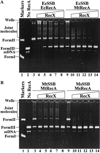
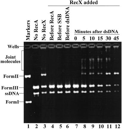
In many eubacteria, coexpression of recX with recA is essential for attenuation of the deleterious effects of recA overexpression; however, the molecular mechanism has remained enigmatic. Here, we show that Mycobacterium tuberculosis RecX binds directly to M. tuberculosis RecA as well as M. smegmatis and E. coli RecA proteins in vivo and in vitro, but not single-stranded DNA binding protein. The direct association of RecX with RecA failed to regulate the specificity or extent of binding of RecA either to DNA or ATP, ligands that are central to activation of its functions. Significantly, RecX severely impeded ATP hydrolysis and the generation of heteroduplex DNA promoted by homologous, as well as heterologous, RecA proteins. These findings reveal a mode of negative regulation of RecA, and imply that RecX might act as an anti-recombinase to quell inappropriate recombinational repair during normal DNA metabolism.
Figures


Similar articles
-
Characterization of DNA strand transfer promoted by Mycobacterium smegmatis RecA reveals functional diversity with Mycobacterium tuberculosis RecA.Biochemistry. 2003 Jun 17;42(23):7216-25. doi: 10.1021/bi0340548. Biochemistry. 2003. PMID: 12795618
-
Characterization of Staphylococcus aureus RecX protein: Molecular insights into negative regulation of RecA protein and implications in HR processes.J Biochem. 2023 Jul 31;174(3):227-237. doi: 10.1093/jb/mvad039. J Biochem. 2023. PMID: 37115499
-
Escherichia coli RecX inhibits RecA recombinase and coprotease activities in vitro and in vivo.J Biol Chem. 2003 Jan 24;278(4):2278-85. doi: 10.1074/jbc.M210496200. Epub 2002 Nov 9. J Biol Chem. 2003. PMID: 12427742
-
RecA and DNA recombination: a review of molecular mechanisms.Biochem Soc Trans. 2019 Oct 31;47(5):1511-1531. doi: 10.1042/BST20190558. Biochem Soc Trans. 2019. PMID: 31654073 Review.
-
The RecA protein as a recombinational repair system.Mol Microbiol. 1991 Jun;5(6):1295-9. doi: 10.1111/j.1365-2958.1991.tb00775.x. Mol Microbiol. 1991. PMID: 1787786 Review.
Cited by
-
RecX facilitates homologous recombination by modulating RecA activities.PLoS Genet. 2012;8(12):e1003126. doi: 10.1371/journal.pgen.1003126. Epub 2012 Dec 20. PLoS Genet. 2012. PMID: 23284295 Free PMC article.
-
Targeting evolution of antibiotic resistance by SOS response inhibition.Comput Struct Biotechnol J. 2021 Jan 11;19:777-783. doi: 10.1016/j.csbj.2021.01.003. eCollection 2021. Comput Struct Biotechnol J. 2021. PMID: 33552448 Free PMC article. Review.
-
Lineage structure of Streptococcus pneumoniae may be driven by immune selection on the groEL heat-shock protein.Sci Rep. 2017 Aug 22;7(1):9023. doi: 10.1038/s41598-017-08990-z. Sci Rep. 2017. PMID: 28831154 Free PMC article.
-
Phylogeny vs genome reshuffling: horizontal gene transfer.Indian J Microbiol. 2008 Jun;48(2):228-42. doi: 10.1007/s12088-008-0034-1. Epub 2008 Jul 27. Indian J Microbiol. 2008. PMID: 23100716 Free PMC article.
-
Cyclic di-AMP regulates genome stability and drug resistance in Mycobacterium through RecA-dependent and RecA-independent recombination.PNAS Nexus. 2024 Dec 12;3(12):pgae555. doi: 10.1093/pnasnexus/pgae555. eCollection 2024 Dec. PNAS Nexus. 2024. PMID: 39697181 Free PMC article.
References
-
- Radding C. M. (1991) J. Biol. Chem. 266, 5355-5358. - PubMed
-
- Cox M. M. (2001) Annu. Rev. Genet. 35, 53-82. - PubMed
-
- Kumar R. A., Vaze, M. B., Chandra, N. R., Vijayan, M. & Muniyappa, K. (1996) Biochemistry 35, 1793-1802. - PubMed
-
- Vaze M. B. & Muniyappa, K. (1999) Biochemistry 38, 3175-3186. - PubMed
Publication types
MeSH terms
Substances
Grants and funding
LinkOut - more resources
Full Text Sources
Molecular Biology Databases
