. 2002 Sep 17;99(19):12339-44.
doi: 10.1073/pnas.192276999. Epub 2002 Sep 6.

Bcrp1 gene expression is required for normal numbers of side population stem cells in mice, and confers relative protection to mitoxantrone in hematopoietic cells in vivo

Affiliations

Bcrp1 gene expression is required for normal numbers of side population stem cells in mice, and confers relative protection to mitoxantrone in hematopoietic cells in vivo

Sheng Zhou et al. Proc Natl Acad Sci U S A. .

Abstract

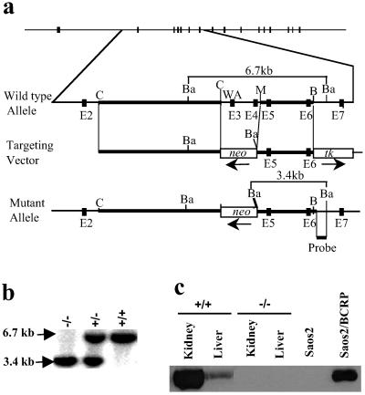
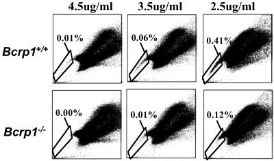
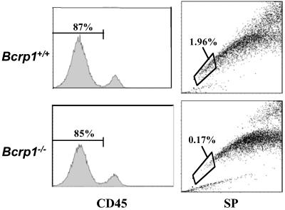
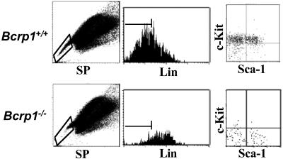
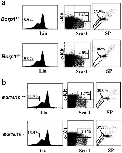
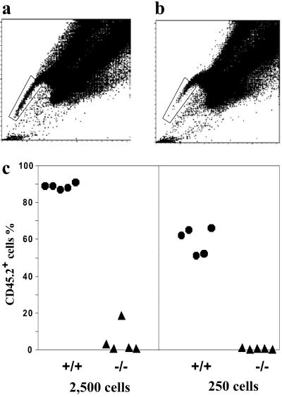
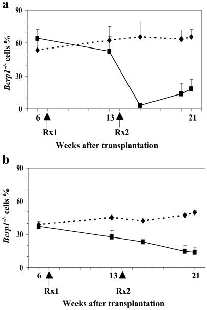
Hematopoietic stem cells (HSCs) can be identified by a "side population" (SP) phenotype. Previous studies have implicated the ATP binding cassette transporter genes Mdr1a/1b and/or Bcrp1 in the SP phenotype. To define the relative role of these transporters, we generated Bcrp1 null mice and evaluated HSCs both functionally and phenotypically. Loss of Bcrp1 gene expression, but not Mdr1a/1b, led to a significant reduction in the number of SP cells in the bone marrow and in skeletal muscle. In the bone marrow, there was a nearly absolute loss of lineage negative, c-Kit-positive, Sca-1-positive SP cells, and the residual SP cells were depleted of repopulating cells in a transplant assay, demonstrating that Bcrp1 expression is necessary for the SP phenotype in HSCs. Furthermore, Bcrp1 null hematopoietic cells were significantly more sensitive to mitoxantrone in drug-treated transplanted mice. These results show that Bcrp1 gene expression alone defines the SP stem cell phenotype, and suggest that the physiological function of Bcrp1 expression in HSCs is to provide protection from cytotoxic substrates.


Figures

Fig 1.
Fig 1.
Targeted disruption of the Bcrp1 locus. (a) The top line shows the 16 exons of Bcrp1 as filled boxes. The wild-type allele is shown in more detail below, with exons 2–7 shown as dark boxes, with restriction sites shown as indicated (C, ClaI; B, BglII; M, MunI; Ba, BamHI). Exon 3 contains the Walker A (WA) motif necessary for Bcrp1 efflux function. The targeting vector was designed to replace the exons 3 and 4 with a pgk-neo cassette. Thick lines show the 5′ and 3′ homologous arms used for recombination, and the location and orientation of the pgk-neo and hsv-tk cassettes are shown. The position of probe for screening correctly targeted ES clones and mice is indicated below the schematic for the mutant allele. The diagnostic fragment sizes for the wild-type (6.7 kb) and mutant allele (3.4 kb) with BamHI digestion is for both alleles. (b) Representative Southern blot analysis of tail DNA from Bcrp1−/−, Bcrp1+/−, and Bcrp1+/+ mice after BamHI digestion. (c) Immunoblot of kidney and liver tissue lysates from wild-type and Bcrp1−/− mice using a polyclonal anti-ABCG2 antibody. As controls, lysates from Saos2 and Saos2 cells expressing BCRP (Saos2/BCRP) are also shown.
Fig 2.
Fig 2.
SP cell analysis of bone marrow cells from Bcrp1−/− mice at Hoechst concentrations of 4.5, 3.5, and 2.5 μg/ml. The results from a wild-type mouse are shown (Upper), as are the results of a Bcrp1−/− mouse (Lower). The percentage of SP cells in the total bone marrow is shown for each sample.
Fig 3.
Fig 3.
SP cell analysis of CD45 skeletal muscle cells from Bcrp1−/− mice. Single cell suspensions from skeletal muscle tissue obtained from wild-type and Bcrp1−/− mice were stained with an anti-CD45 antibody and Hoechst dye. The CD45, nonhematopoietic cells were gated (Left) and analyzed for SP cells (Right). The proportion of SP cells within the CD45 gate is shown for each sample.
Fig 4.
Fig 4.
Immunophenotype of SP cells from Bcrp1−/− mice. Bone marrow cells from wild-type (Upper) and Bcrp1−/− mice (Lower) were stained with 2.5 μg/ml of Hoechst dye and antibodies to lineage markers, Sca-1 and c-Kit. Cells were first gated for SP cells (Left), and then analyzed for proportion of Lin cells in the SP gate (Center). (Right) The distribution of c-Kit and Sca-1 markers in the gated SP, Lin cells.
Fig 5.
Fig 5.
Analysis of K+S+L cells in Bcrp1−/− and Mdr1a/1b−/− mice. (a) Bone marrow cells from wild-type (Upper) and Bcrp1−/− mice (Lower) were stained with Hoechst dye and antibodies to Lin, Sca-1, and c-Kit. (Left) The percentage of Lin cells in the bulk population. (Center) The percentage of c-Kit- and Sca-1-positive cells in the gated Lin population. (Right) The proportion of SP cells in gated K+S+L cells. (b) The same analysis was performed on wild-type mice (Upper) and Mdr1a/1b knockout mice (Lower). Gating and cell percentages were done as described above.
Fig 6.
Fig 6.
Repopulation capability of sorted SP cells from Bcrp1−/− bone marrow. Sorting gate for wild-type SP cells (a) and Bcrp1−/− SP cells (b). (c) Reconstitution level of peripheral blood myeloid cells derived from the sorted SP cells 4 weeks after transplantation. The number of SP cells transplanted into each mouse is indicated at the bottom. Wild-type SP cells (•), Bcrp1−/− SP cells (▴).
Fig 7.
Fig 7.
Effects of mitoxantrone on the number of Bcrp1−/− hematopoietic cells in the peripheral blood. (a) Bcrp1−/− chimerism in granulocytes (Gr-1+) and monocytes (Mac-1+). (b) Bcrp1−/− chimerism in B lymphocytes (B220+) and T lymphocytes (Thy1.2+). Mitoxantrone treated group (▪), untreated control group (⧫), n = 3. Rx1: first round of treatment with 1 mg/kg mitoxantrone; Rx2: second round of treatment with 2 mg/kg mitoxantrone.

References

    1. Spangrude G. J. & Johnson, G. R. (1990) Proc. Natl. Acad. Sci. USA 87, 7433-7437. - PMC - PubMed
    1. Wolf N. S., Kone, A., Priestley, G. V. & Bartelmez, S. H. (1993) Exp. Hematol. 21, 614-622. - PubMed
    1. Goodell M. A., Brose, K., Paradis, G., Conner, A. S. & Mulligan, R. C. (1996) J. Exp. Med. 183, 1797-1806. - PMC - PubMed
    1. Goodell M. A., Rosenzweig, M., Kim, H., Marks, D. F., DeMaria, M., Paradis, G., Grupp, S. A., Sieff, C. A., Mulligan, R. C. & Johnson, R. P. (1997) Nat. Med. 3, 1337-1345. - PubMed
    1. Storms R. W., Goodell, M. A., Fisher, A., Mulligan, R. C. & Smith, C. (2000) Blood 96, 2125-2133. - PubMed

Publication types

MeSH terms

Substances