. 2002 Sep 17;99(19):12327-32.
doi: 10.1073/pnas.192301399. Epub 2002 Sep 6.

Induction of cyclooxygenase-2 in a mouse model of Peutz-Jeghers polyposis

Affiliations

Induction of cyclooxygenase-2 in a mouse model of Peutz-Jeghers polyposis

Derrick J Rossi et al. Proc Natl Acad Sci U S A. .

Abstract

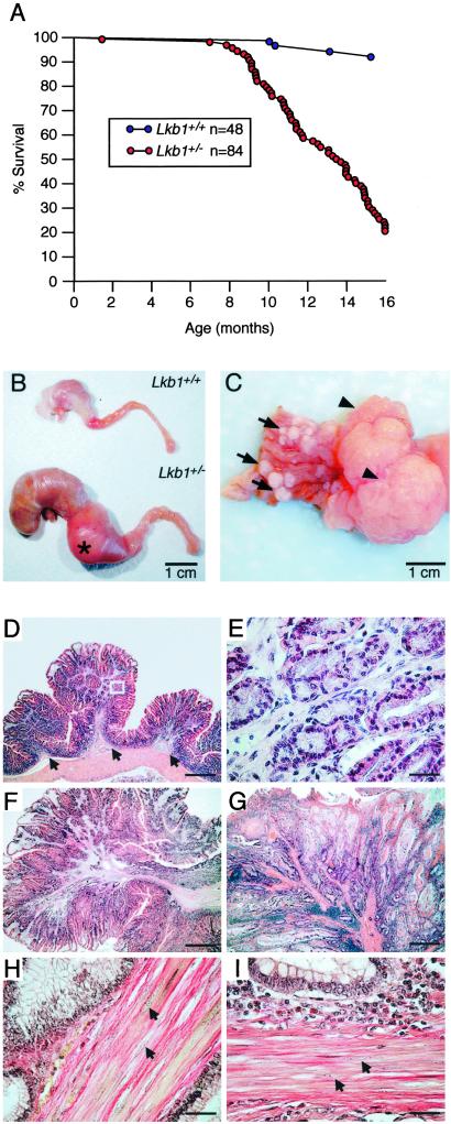
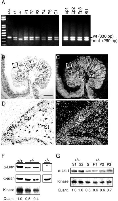
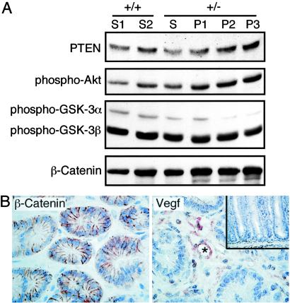
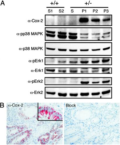
Inactivating germ-line mutations of LKB1 lead to Peutz-Jeghers syndrome (PJS). We have generated mice heterozygous for a targeted inactivating allele of Lkb1 and found that they develop severe gastrointestinal polyposis. In all cases, the polyps arising in the Lkb1+/- mice were found to be hamartomas that were histologically indistinguishable from polyps resected from PJS patients, indicating that Lkb1+/- mice model human PJS polyposis. No evidence for inactivation of the remaining wild-type Lkb1 allele in Lkb1+/- -associated polyps was observed. Moreover, polyps and other tissues in heterozygote animals exhibited reduced Lkb1 levels and activity, indicating that Lkb1 was haploinsufficient for tumor suppression. Analysis of the molecular mechanisms characterizing Lkb1+/- polyposis revealed that cyclooxygenase-2 (COX-2) was highly up-regulated in murine polyps concomitantly with activation of the extracellular signal-regulated kinases 1 and 2 (Erk1/2). Subsequent examination of a large series of human PJS polyps revealed that COX-2 was also highly up-regulated in the majority of these polyps. These findings thereby identify COX-2 as a potential target for chemoprevention in PJS patients.


Figures

Fig 1.
Fig 1.
Increased mortality and polyposis modeling PJS in Lkb1+/− mice. (A) Survival curve of 48 wt (blue points) and 84 Lkb1+/− mice (red points) with points representing animals that died or were killed because of ill health. (B) Stomach, duodenum, and proximal jejunum of Lkb1+/− and Lkb1+/+ littermate. (C) Representative gastric stomach of an Lkb1+/− animal showing multiple small polyps (arrows) and large pyloric junction polyps (arrowheads). (D) Hematoxylin/eosin staining of a cross-section of glandular stomach from a Lkb1+/− mouse showing three small hamartomatous polyps. Note the smooth muscle core that is contiguous with the muscularis mucosa (arrows). (E) Higher magnification of the boxed region in D. (F and G) Hematoxylin/eosin staining of a murine polyp (F) and human PJS patient polyp (G). (H and I) Herovici's staining of a murine polyp (H) and a human PJS patient polyp (I) with smooth muscle nuclei indicated with arrows (compare to H). (Bars in D, F, and G = 0.8 mm; bars in E, H, and I = 40 μm.)
Fig 2.
Fig 2.
Lkb1 is haploinsufficient for tumor suppression. (A) PCR genotyping of Lkb1 from DNAs extracted from five polyps (P1–P5), one control Lkb1+/− tail (C1), and yolk sacs dissected from wt (+/+), heterozygous (+/−), and Lkb1 null (−/−) E9.5 embryos (Left), and of laser-microdissected epithelial (Ep1–Ep3) or stromal (St) cells from polyps of multiple Lkb1+/− animals (Right). A 330-bp wt band and a 260-bp mutant band are indicated. (B and C) Lkb1 in situ hybridization showing brightfield (B) and darkfield (C) images of a polyp and adjacent unaffected gastric epithelia from a Lkb1+/− animal. (D and E) Higher magnification of the brightfield (D) and darkfield (E) images of boxed regions shown in B and C demonstrating Lkb1 mRNA expression both in epithelial [Ep] and stromal [St] cell types. (F) Western blotting analysis of lysates from wt (+/+) or Lkb1 heterozygous (+/−) MEF cultures or Lkb1 null whole embryos (−/−) immunoblotted for Lkb1 (α-Lkb1) and actin (α-actin), and kinase assay showing Lkb1 autocatalytic activity with quantitation (Quant.). (G) Western blotting analysis of lysates from two independently isolated wt (+/+) stomachs (S1 and S2), and Lkb1 heterozygous (+/−) unaffected stomach (S) and three individual polyps (P1–P3) with α-Lkb1 (Upper), and Lkb1 kinase assay with quantitation (Quant.) (Lower). (Bar = 0.7 mm.)
Fig 3.
Fig 3.
Characterization of Lkb1+/− polyposis. (A) Western blot analysis of lysates from two independent wt (+/+) stomachs (S1 and S2), Lkb1 heterozygous (+/−) unaffected stomach (S), and three individual polyps (P1–P3) with Abs specific for PTEN, phospho-Akt (Ser-473), phospho-GSK-3α/3β (Ser-21/Ser-9), and β-catenin. (B) Immunostaining of β-catenin (Left) and VEGF (Right) of polyp from Lkb1+/− animals at ×400 magnification. (Inset) Adjacent unaffected gastric mucosa immunostained for VEGF at ×200 magnification. The blood vessel (Right) is marked by an asterisk (*).
Fig 4.
Fig 4.
COX-2 induction in murine Lkb1+/− and PJS polyposis. (A) Western blot analysis of lysates from two independent wt (+/+) stomachs (S1 and S2), Lkb1 heterozygous (+/−) unaffected stomach (S), and three individual polyps (P1–P3) with Abs specific for COX-2, activated p38 MAPK (pp38 MAPK), p38 MAPK (p38 MAPK), activated Erk1 kinase (pErk1), Erk1 kinase (Erk1), activated Erk2 kinase (pErk2), and Erk2 kinase (Erk2). (B) COX-2 immunostaining of a small intestinal polyp from patient PJ8 showing epithelial cytoplasmic localization (Left) with high magnification (Inset), and a serial section after Ab preadsorption with a COX-2 peptide (Right).

References

    1. Peutz J. L. A. (1921) Ned. Maandsch. Geneeskd. 10, 134-146.
    1. Jeghers H., McKusick, V. A. & Katz, K. H. (1949) N. Engl. J. Med. 241, 1031-1036. - PubMed
    1. Vasen H. F. (2000) J. Clin. Oncol. 18, 81S-92S. - PubMed
    1. Giardiello F. M., Welsh, S. B., Hamilton, S. R., Offerhaus, G. J., Gittelsohn, A. M., Booker, S. V., Krush, A. J., Yardley, J. H. & Luk, G. D. (1987) N. Engl. J. Med. 316, 1511-1514. - PubMed
    1. Boardman L. A., Thibodeau, S. N., Schaid, D. J., Lindor, N. M., McDonnell, S. K., Burgart, L. J., Ahlquist, D. A., Podratz, K. C., Pittelkow, M. & Hartmann, L. C. (1998) Ann. Intern. Med. 128, 896-899. - PubMed

Publication types

MeSH terms