. 2002 Oct 1;99(20):12663-8.
doi: 10.1073/pnas.192445599. Epub 2002 Sep 6.

The hDcp2 protein is a mammalian mRNA decapping enzyme

Affiliations

The hDcp2 protein is a mammalian mRNA decapping enzyme

Zuoren Wang et al. Proc Natl Acad Sci U S A. .

Abstract

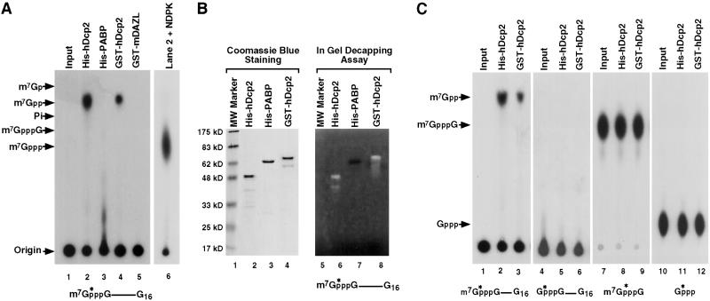
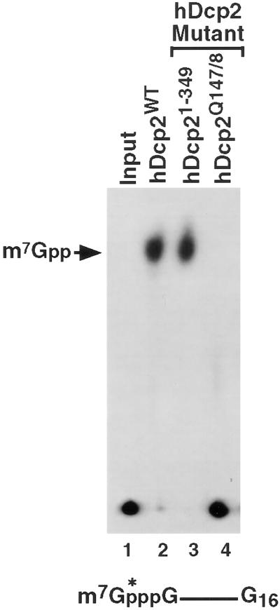
Decapping of mRNA is a critical step in eukaryotic mRNA turnover, yet the proteins involved in this activity remain elusive in mammals. We identified the human Dcp2 protein (hDcp2) as an enzyme containing intrinsic decapping activity. hDcp2 specifically hydrolyzed methylated capped RNA to release m(7)GDP; however, it did not function on the cap structure alone. hDcp2 is therefore functionally distinct from the recently identified mammalian scavenger decapping enzyme, DcpS. hDcp2-mediated decapping required a functional Nudix (nucleotide diphosphate linked to an X moiety) pyrophosphatase motif as mutations in conserved amino acids within this motif disrupted the decapping activity. hDcp2 is detected exclusively in the cytoplasm and predominantly cosediments with polysomes. Consistent with the localization of hDcp2, endogenous Dcp2-like decapping activity was detected in polysomal fractions prepared from mammalian cells. Similar to decapping in yeast, the presence of the poly(A) tail was inhibitory to the endogenous decapping activity, yet unlike yeast, competition of cap-binding proteins by cap analog did not influence the efficiency of decapping. Therefore the mammalian homologue of the yeast Dcp2 protein is an mRNA decapping enzyme demonstrated to contain intrinsic decapping activity.


Figures

Figure 1
Figure 1
Schematic of Dcp2 proteins from different organisms. Dcp2 proteins from diverse species are schematically represented and aligned by clustalw relative to the Nudix motif. Three distinct regions of high homology are noted. An N-terminal conserved 38-aa box A, a 109-aa Nudix fold with the central 23-aa Nudix motif region, and a 20-aa box B segment are indicated (see Fig. 5). The aa position of the conserved regions within each protein is denoted above the protein schematics and the total number of amino acids in each protein is shown on the right. The degree of identity within the Nudix motif among the different Dcp2 proteins relative to the human protein is as follows: 98% with mouse (GenBank accession no. AK017809); 44% with Caenorhabditis elegans (GenBank accession no. NM_070208), 53% with Drosophila melanogaster (GenBank accession no. AE003529), 47% with Schizosaccharomyces pombe (GenBank accession no. CAB11648), and 38% with Saccharomyces cerevisiae (GenBank accession no. P53550).
Figure 2
Figure 2
Recombinant hDcp2 contains intrinsic decapping activity specific to methylated capped RNA. (A) In vitro decay assays were carried out by incubating 0.5 μg of the indicated histidine-tagged (His-hDcp2 and His-PABP) or GST-fused (GST-hDcp2 and GST-mDAZL) recombinant proteins with cap-labeled pcP-G16 RNA at 37°C for 30 min. Reaction products were resolved by polyethyleneimine-TLC developed in 0.75 M LiCl. An aliquot of the reaction product used in lane 2 was treated with NDPK and revolved by TLC (lane 6) to confirm the original product was m7GDP. Standards were simultaneously developed and their positions are denoted on the left. The substrate used in the assay is schematically denoted at the bottom. The asterisks represent the position of the 32P-labeling and the line denotes the RNA. (B) One microgram of each recombinant protein was separated either on a standard SDS/PAGE (Left) or an identical gel containing 32P-cap-labeled pcP-G16 RNA polymerized into the gel (Right). Migration of the proteins was visualized by Coomassie blue staining (Left). An in-gel decapping activity assay was carried out with the gel containing 32P-labeled-capped RNA as described in Materials and Methods, and an autoradiography of the gel is shown (Right). Decapping of the RNA produces a clearing of the radioactive background upon release of the labeled cap substrate that diffuses out of the gel. Both the His-hDcp2 (lane 6) and GST-hDcp2 (lane 8) are capable of decapping whereas the His-PABP control (lane 7) was not. Protein size markers are shown on the left. (C) In vitro decay assays were carried out similar to A but used different substrates. Cap-labeled RNA lacking the N-7 methyl moiety was used in lanes 4–6, labeled methylated cap analog dinucleotide in lanes 7–9, and [α-32P]GTP in lanes 10–12. hDcp2 efficiently hydrolyzed methylated cap-labeled RNA (lanes 2 and 3) but not the other three substrates. The input substrates are shown in lanes 1, 4, 7, and 10. The recombinant protein used for the assays are indicated at the top.
Figure 3
Figure 3
Significance of the hDcp2 Nudix motif in decapping. In vitro decay assays were carried out by using cap-labeled RNA and 0.5 μg of the indicated recombinant protein at 37°C for 30 min. Wild-type hDcp2 protein, the truncated protein lacking the C-terminal 71 aa (hDcp21–349), and the double point mutation substituting glutamine for glutamic acid at amino acids 147 and 148 (hDcp2Q147/8) are indicated. The resulting decapping products were resolved on TLC plates and the labeling is as described in the legend to Fig. 1.
Figure 4
Figure 4
Endogenous decapping activity cosediments with polysomes. (A) Western analysis using affinity-purified antibody directed against hDcp2 was carried out. Fifty nanograms of His-hDcp2 was used in lanes 1, and extract derived from 1 × 10⁶ K562 cells was fractionated and resolved in lanes 2–7 as indicated. The protein size markers are shown on the left. (B) An in vitro decay assay was carried out by incubating cap-labeled pcP-G16 RNA with 30 μg of the indicated K562 cell extract in the absence or presence of 100 μM cap analog. The obtained products were resolved by TLC as shown in lanes 2–9. The products of lanes 5–9 were treated with NDPK and then resolved by TLC (lanes 10–14). (C) In vitro decay assay was carried out by incubating 30 μg of K562 RSW fraction with cap-labeled pcP-G16 RNA or pcP-G16-A60 RNA in the absence or presence of 500 ng of cold poly(A) competitor at 37°C for 30 min. The obtained products were resolved by TLC as shown in lanes 2, 3, 5, and 6.

Comment in

References

    1. Beelman C A, Stevens A, Caponigro G, LaGrandeur T E, Hatfield L, Fortner D M, Parker R. Nature (London) 1996;382:642–646. - PubMed
    1. LaGrandeur T E, Parker R. EMBO J. 1998;17:1487–1496. - PMC - PubMed
    1. Larimer F W, Hsu C L, Maupin M K, Stevens A. Gene. 1992;120:51–57. - PubMed
    1. Muhlrad D, Decker C J, Parker R. Genes Dev. 1994;8:855–866. - PubMed
    1. Czaplinski K, Ruiz-Echevarria M J, Gonzalez C I, Peltz S W. BioEssays. 1999;21:685–696. - PubMed

Publication types

MeSH terms

Associated data