

doi: 10.1046/j.1469-7580.2002.00083.x.
A quantitative analysis of perineurial cell basement membrane collagen IV, laminin and fibronectin in diabetic and non-diabetic human sural nerve
Affiliations
- PMID: 12220126
- PMCID: PMC1570905
- DOI: 10.1046/j.1469-7580.2002.00083.x
Item in Clipboard
A quantitative analysis of perineurial cell basement membrane collagen IV, laminin and fibronectin in diabetic and non-diabetic human sural nerve
J Anat.
2002 Aug.
Abstract
The thickness of the perineurial cell basement membrane was examined in diabetic and non-diabetic human sural nerve. A significant increase in thickness was found in the diabetic group. The nature of this thickening was investigated using immunohistochemistry and image analysis in order to semi-quantify three of the major intrinsic components of the perineurial cell basement membrane: collagen IV, laminin and fibronectin. Amounts of all three components were shown to be increased in the diabetic group, but not significantly so. However, significant linear correlations between fascicle size and perineurial collagen IV, laminin and fibronectin were identified in both diabetic and non-diabetic nerve.
Figures
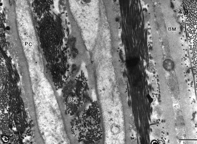
Part of the perineurium of a human sural nerve. Each perineurial cell (PC) is bordered on both surfaces by basement membrane (BM). Between each perineurial cell layer are collagen fibrils (CF). Scale bar = 1 μm.
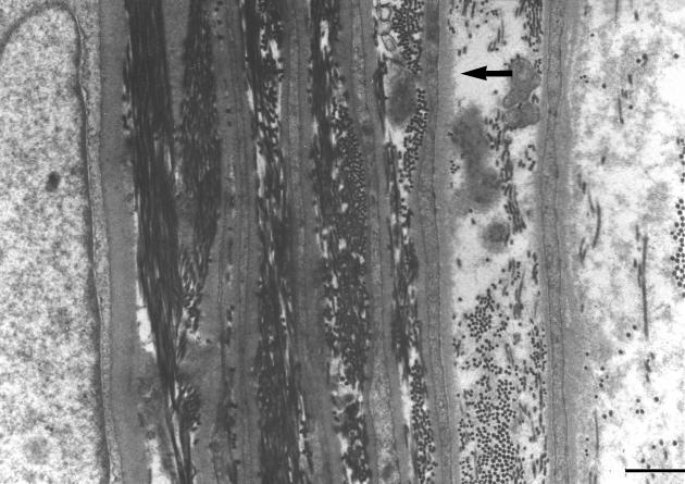
Part of an electron micrograph montage of the perineurium from which basement membrane thickness measurements were made. Measurements were taken at 2-cm intervals along the length of each perineurial cell basement membrane (arrow). Scale bar = 1 μm.
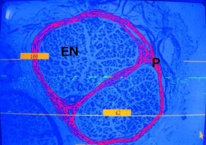
A single human nerve fascicle thresholded for assessment of perineurial collagen IV content, endoneurium (EN), perineurium (P). Highlighted pixels representing collagen IV found elsewhere in the nerve structure have been excluded. (×100).
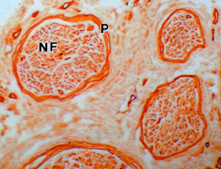
A paraffin wax section of a human sural nerve immunostained for collagen IV. The perineurium (P) shows deep staining. Complete nerve fascicles (NF) were easily visualized with the image analyser using a low-power objective (×100).
Similar articles
-
Changes in steady-state levels of mRNAs coding for type IV collagen, laminin and fibronectin following capillary basement membrane thickening in human adult onset diabetes.Connect Tissue Res. 1990;25(1):77-85. doi: 10.3109/03008209009009814. Connect Tissue Res. 1990. PMID: 2245601
-
The extracellular matrix of peripheral nerve in diabetic polyneuropathy.Acta Neuropathol. 2000 May;99(5):539-46. doi: 10.1007/s004010051158. Acta Neuropathol. 2000. PMID: 10805099
-
Relationship between fascicle size and perineurial collagen IV content in diabetic and control human peripheral nerve.Histopathology. 2000 Jun;36(6):551-5. doi: 10.1046/j.1365-2559.2000.00897.x. Histopathology. 2000. PMID: 10849098
-
Comparison of various basement membrane components in benign and malignant peripheral nerve tumours.Virchows Arch A Pathol Anat Histopathol. 1992;421(4):331-8. doi: 10.1007/BF01660980. Virchows Arch A Pathol Anat Histopathol. 1992. PMID: 1413494
-
Basement membrane components are key players in specialized extracellular matrices.Cell Mol Life Sci. 2010 Sep;67(17):2879-95. doi: 10.1007/s00018-010-0367-x. Epub 2010 Apr 29. Cell Mol Life Sci. 2010. PMID: 20428923 Free PMC article. Review.
Cited by
-
Effect of Hesperidin on Sciatic Nerve Damage in STZ-Induced Diabetic Neuropathy: Modulation of TRPM2 Channel.Neurotox Res. 2023 Dec;41(6):638-647. doi: 10.1007/s12640-023-00657-0. Epub 2023 Jul 13. Neurotox Res. 2023. PMID: 37439953
-
Case report: Intraneural perineurioma in dogs: a case series and brief literature review.Front Vet Sci. 2024 Jan 11;10:1233230. doi: 10.3389/fvets.2023.1233230. eCollection 2023. Front Vet Sci. 2024. PMID: 38274660 Free PMC article.
-
Benfotiamine reduced collagen IV contents of sciatic nerve in hyperglycemic rats.J Diabetes Metab Disord. 2021 Feb 20;20(1):21-30. doi: 10.1007/s40200-020-00666-2. eCollection 2021 Jun. J Diabetes Metab Disord. 2021. PMID: 34222057 Free PMC article.
-
Diabetic nephropathy and extracellular matrix.J Histochem Cytochem. 2012 Dec;60(12):976-86. doi: 10.1369/0022155412465073. Epub 2012 Oct 27. J Histochem Cytochem. 2012. PMID: 23103723 Free PMC article. Review.
-
Correlation between diffusion tensor indices and fascicular morphometric parameters of peripheral nerve.Front Physiol. 2023 Feb 23;14:1070227. doi: 10.3389/fphys.2023.1070227. eCollection 2023. Front Physiol. 2023. PMID: 36909220 Free PMC article.
References
-
- Adler S, Anderson P, Xu G, Ihm C, Nast C, Gullermo R, Glassock R. Early renal procollagen ×1 (IV) gene expression in diabetic rats occurs in deep to superficial cortical glomeruli (Abstract) Clin. Res. 1989;37:484A.
-
- Bradley JL, Thomas PK, King RMH, Watkins PJ. A comparison of perineurial and vascular basal laminal changes in diabetic neuropathy. Acta Neuropathol. 1994;88:426–432. - PubMed
-
- Bradley JL, King RHM, Muddle JR, Thomas PK. The extracellular matrix of peripheral nerve in diabetic polyneuropathy. Acta Neuropath. 2000;99:539–546. - PubMed
-
- Brownlee M, Spiro RG. Glomerular basement membrane metabolism in the diabetic rat. In vivo studies. Diabetes. 1979;28:121–125. - PubMed
MeSH terms
Substances
LinkOut - more resources
Full Text Sources
Medical
