Review
. 2002 Oct;25(10):518-24.
doi: 10.1016/s0166-2236(02)02241-5.

Stressed-out, or in (utero)?

Affiliations
Review

Stressed-out, or in (utero)?

Sarit Avishai-Eliner et al. Trends Neurosci. 2002 Oct.

Abstract

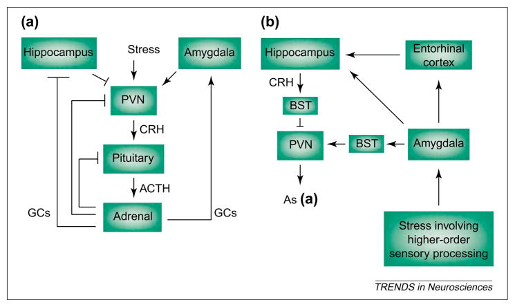
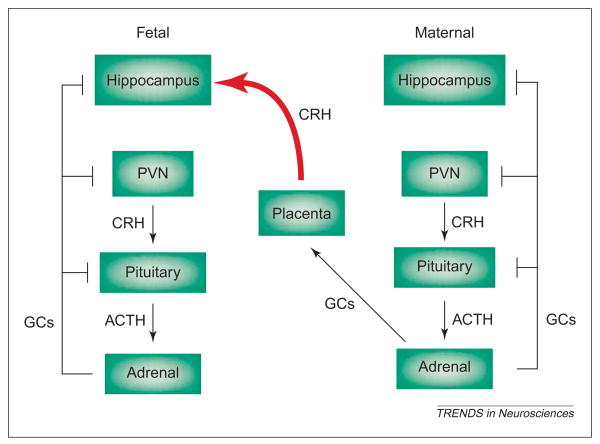
The molecular and cellular mechanisms by which plasticity is induced in the mature CNS (and, specifically, in the hippocampus) by environmental input are progressively being elucidated. However, the mechanisms - and even the existence - of functional and structural effects of environmental input (and, particularly, stress) early in life are incompletely understood. Here, we discuss recent evidence that stressful stimuli have a significant impact on neonatal (rat) and prenatal (human) hippocampal function and integrity. Stressful signals provoke expression and release of neuromodulators, including the peptide corticotropin-releasing hormone (CRH), leading to activation of CRH receptors on principal hippocampal neurons. Although physiological activation of these receptors promotes synaptic efficacy, pathological levels of CRH at hippocampal synapses contribute to neuronal death. Thus, early-life stress could constitute a 'double-edged sword': mild stress might promote hippocampal-dependent cognitive function, whereas severe stress might impair neuronal function and survival, both immediately and in the long-term. Importantly, these CRH-mediated processes could be targets of preventive and interventional strategies.


Figures

Fig. 1
Fig. 1
Stress-activated pathways include the neuroendocrine hypothalamic–pituitary–adrenal axis (a) and the central, limbic stress-loop (b). (a) ‘Physiological’ stress signals reach the hypothalamus, causing secretion of corticotropin-releasing hormone (CRH) from neurons of the paraventricular nucleus (PVN). CRH induces release of adrenocorticotropic hormone (ACTH) from the pituitary, and ACTH elicits secretion of glucocorticoids (GCs) from the adrenal gland. GCs cross the blood–brain barrier and activate specific receptors in hippocampus (and other CNS regions) to ‘shut off’ the neuroendocrine stress response. By contrast, GCs increase CRH mRNA expression in the amygdala, facilitating stress-responses [19,35], whereas pituitary ACTH reduces CRH mRNA levels in the amygdala by direct activation of melanocortin receptors [55]. (b) Stress involving higher-order sensory processing (i.e. with ‘cognitive’ and/or ‘emotional’ aspects) activates limbic pathways constituting the more recently elucidated ‘central’ stress circuit. Stressful stimuli reach the key processor, the central nucleus of the amygdala (ACe), activating the numerous CRH-producing neurons in this region. Locally released CRH acts on cognate receptors on projection neurons of the amygdala, which convey stress-related information (directly, or indirectly via the entorhinal cortex) to the hippocampal formation. Within the hippocampus, stress-induced release of CRH from interneurons in the CA3 and CA1 pyramidal-cell layers enhances synaptic efficacy and influences memory function. Arrows indicate facilitatory projections but do not imply monosynaptic connections. Blunt-ended lines denote inhibitory feedback loops. Abbreviation: BST, bed nucleus of the stria terminalis.
Fig. 2
Fig. 2
Reproducing the effects of stress, administration of corticotropin-releasing hormone (CRH) to immature (10-day-old) rat induces production of Fos protein (black reaction product; arrows) in select populations of neurons in the amygdala (a,b) and hippocampus (c) that bear the CRF1 subtype of CRH receptor (brown immunoreactivity). Coronal sections from animals perfused 2 h after CRH treatment were double-labeled for Fos and CRF1 using standard methods [37,40,61]. CRH induced Fos production (arrows) preferentially in the central nucleus of the amygdala (ACe) (a, b) and in CA3a hippocampal pyramidal cell layer (c). Higher magnification (b) demonstrates cells of the amygdala double-labeled for the nuclear protein Fos and the membranous CRF1. Scale bar, 100 μm for (a,c), 20 μm for (b). Abbreviation: BL, Basolateral nucleus of the amygdala.
Fig. 3
Fig. 3
CRH-positive neuronal populations are abundant in the amygdala (a) and hippocampus (b, c) of the developing (10-day old) rat. Within the amygdala complex, CRH-immunoreactive cell bodies and fibers populate preferentially the central nucleus of the amygdala (ACe), which is involved in ‘processing’ of stress signals [29,35]. In the hippocampus, corticotropin-releasing hormone (CRH)-producing interneurons reside throughout CA1 and CA3 pyramidal-cell layers [stratum pyramidale (SP); b; arrowheads]. (c) A high magnification of the CA1 subfield demonstrates that CRH-immunoreactive interneurons and axon terminals (arrows) richly innervate pyramidal cell somata, presumably influencing the activity of these neurons. Scale bar, 150 μm for (a,b), 25 μm for (c).
Fig. 4
Fig. 4
In the prenatal human, corticotropin-releasing hormone (CRH) derived from maternal placenta could influence the fetal hippocampus. Sustained stress during pregnancy activates the maternal neuroendocrine stress axis, resulting in increased production and release of placental CRH into the bloodstream. In contrast to hypothalamic CRH production, which is suppressed by stress-induced glucocorticoids (GCs), CRH-gene expression in placenta is enhanced by GCs, so that maternal stress leads to progressively higher fetal plasma CRH levels. This maternal-origin CRH reaches the fetal brain (red curved arrow) [54], influencing fetal learning and/or memory functions [12,53], presumably by activating hippocampal CRH receptors. Arrows indicate facilitatory pathways but do not imply monosynaptic connections. Blunt-ended lines denote inhibitory feedback loops.

Similar articles

Cited by

References

    1. Dobbing J, Sands J. Comparative aspects of the brain growth spurt. Early Hum Dev. 1979;3:79–83. - PubMed
    1. Gottlieb A, et al. Rodent brain growth stages: an analytical review. Biol Neonate. 1977;32:166–176. - PubMed
    1. Clancy B, et al. Translating developmental time across mammalian species. Neuroscience. 2001;105:7–17. - PubMed
    1. Herschkowitz N, et al. Neurobiological bases of behavioral development in the first year. Neuropediatrics. 1997;28:296–306. - PubMed
    1. Seress L. Morphological changes in the human hippocampal formation from midgestation to early childhood. In: Nelson CA, Luciana M, editors. Handbook of Developmental Cognitive Neuroscience. MIT Press; 2001. pp. 45–58.

Publication types

Substances