. 2002 Sep 16;158(6):1017-27.
doi: 10.1083/jcb.200205124. Epub 2002 Sep 9.

The role of the Tim8p-Tim13p complex in a conserved import pathway for mitochondrial polytopic inner membrane proteins

Affiliations

The role of the Tim8p-Tim13p complex in a conserved import pathway for mitochondrial polytopic inner membrane proteins

Sean P Curran et al. J Cell Biol. .

Abstract

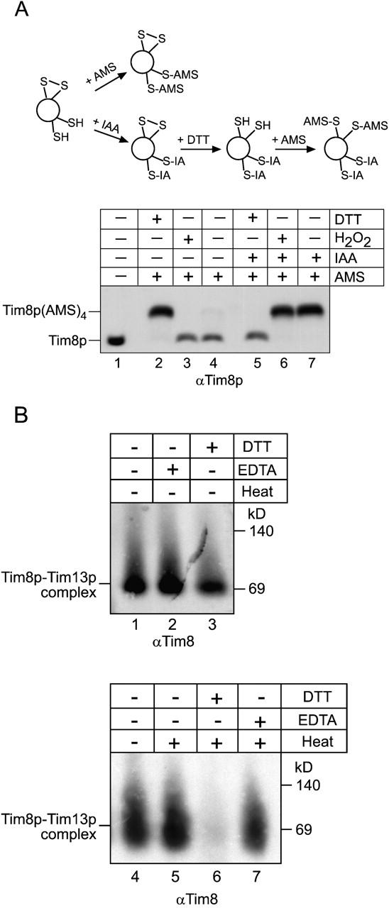
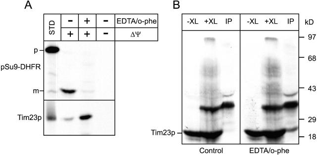
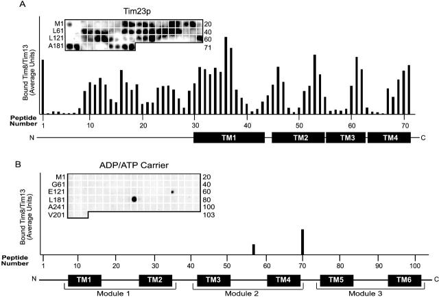
Tim23p is imported via the TIM (translocase of inner membrane)22 pathway for mitochondrial inner membrane proteins. In contrast to precursors with an NH2-terminal targeting presequence that are imported in a linear NH2-terminal manner, we show that Tim23p crosses the outer membrane as a loop before inserting into the inner membrane. The Tim8p-Tim13p complex facilitates translocation across the intermembrane space by binding to the membrane spanning domains as shown by Tim23p peptide scans with the purified Tim8p-Tim13p complex and crosslinking studies with Tim23p fusion constructs. The interaction between Tim23p and the Tim8p-Tim13p complex is not dependent on zinc, and the purified Tim8p-Tim13p complex does not coordinate zinc in the conserved twin CX3C motif. Instead, the cysteine residues seemingly form intramolecular disulfide linkages. Given that proteins of the mitochondrial carrier family also pass through the TOM (translocase of outer membrane) complex as a loop, our study suggests that this translocation mechanism may be conserved. Thus, polytopic inner membrane proteins, which lack an NH2-terminal targeting sequence, pass through the TOM complex as a loop followed by binding of the small Tim proteins to the hydrophobic membrane spanning domains.


Figures

Figure 1.
Figure 1.
Tim23p passes through the TOM complex as a loop and is inserted into the inner membrane. (A) Diagram of Tim23p-DHFR fusions constructed to investigate the import pathway of Tim23p. DHFR–Tim23p, DHFR fused at NH2 terminus of Tim23p; Tim23p–DHFR, DHFR fused to COOH terminus of Tim23p; DHFR–Tim23p–DHFR, DHFR fused to both the NH2- and COOH-termini of Tim23p. (B) Radiolabeled Tim23p and fusion constructs listed in A were synthesized in vitro and incubated with wild-type mitochondria in the absence of a Δψ and in the presence of methotrexate. Half of the import reaction was treated with 5 μg/ml proteinase K (protease) to ensure that the precursor arrested across the outer membrane. Subsequently, the chemical crosslinker DSS was added at 1 mM. After quenching, the sample was denatured and immunoprecipitated with antibodies against Tim8p (IP). Bound proteins were eluted with SDS-containing sample buffer and analyzed by SDS-PAGE and fluorography. STD, 5% of the radioactive prescursor added to each assay. The arrow denotes the location of the monomeric precursor. (C) The radiolabeled precursors in A were imported into wild-type mitochondria at 25°C in the presence or absence of a Δψ. The mitochondria were lysed by osmotic shock at 4°C in the presence of 20 μg/ml proteinase K, followed by incubation with 1 mM PMSF, and extraction with 0.1 M Na2CO3 (pH 11.0). Tim23f, 14 kD COOH-terminal protease-resistant fragment that is inserted into the inner membrane.
Figure 2.
Figure 2.
Tim8p and Tim13p assemble into a 70-kD complex when coexpressed in E. coli. (A) An expression plasmid was constructed in which TIM8 and TIM13 were each placed behind an E. coli ribosomal binding site. After transformation into E. coli host BL21(DE3) and induction, the Tim8p–Tim13p complex was purified using column chromatography (Materials and methods). The fractions from the final purification step (Superose 12 column) were analyzed for Tim8p and Tim13p by SDS-PAGE and Coomassie staining. Tim8p and Tim13p are denoted by the arrow. (B) Fraction 12 (as shown in A) and a mitochondrial intermembrane space sample (IMS) prepared by osmotic shock were analyzed on a 6–16% blue native gel followed by immunoblotting with antiserum for Tim8p. Proteins were identified by incubation with [125I]-protein A. (C) The recombinant Tim8p–Tim13p complex and a mitochondrial IMS were separated on a 6.5% native gel. Immunoblot analysis was performed with monospecific antiserum against Tim8p (αTim8) and Tim13p (αTim13) and detected with [125I]-protein A.
Figure 3.
Figure 3.
The recombinant and native Tim8p–Tim13p complexes have identical properties. (A) For intermolecular crosslinking assays, an intermembrane space sample and the recombinant Tim8p–Tim13p complex were incubated with 0.1% glutaraldehyde (XL) and aliquots were removed at the indicated time points. Crosslinked products were separated on 16% Tricine gels followed by immunoblot analysis with monospecific antisera against Tim8p (αTim8). The number of subunits is indicated at the right. Electrophoretic mobility (lowest panel) of the products from the 10-min time point of crosslinking was plotted against the logarithm of Mw/Mm, where Mw is the relative molecular mass of the crosslinked species and Mm is the average molecular mass of the Tim8p or Tim13p monomer (11 kD). (B) The recombinant Tim8p–Tim13p complex and a mitochondrial intermembrane space sample were incubated with 5 μg/ml trypsin at 37°C. At the indicated periods, aliquots were removed and soybean trypsin inhibitor was added. Samples were separated by SDS-PAGE followed by immunoblot analysis with antibodies for Tim8p or Tim13p. Protease resistance (% Res) was quantified by scanning laser densitometry and the zero time point was set at 100%.
Figure 4.
Figure 4.
CD studies on the purified Tim8p–Tim13p complex. The CD spectra of the recombinant Tim8p–Tim13p complex at (a) 4°C, (b) 95°C, and (c) 4°C after heating to 95°C. Each spectrum was an average of eight scans. The secondary structure of the spectra at 4°C (curve a) before and after thermal denaturation (curve c) was predicted by a self-consistent method and the convex constraint algorithm (Perczel et al., 1992; Sreerama and Woody, 1993) (bottom).
Figure 5.
Figure 5.
The Tim8p–Tim13p complex does not require Zn2 + for structural stability. (A) The sulfhydryl groups on the cysteines residues of the twin CX3C motif are occupied. A thiol-trapping method (Jakob et al., 1999) was used to assess the state of the cysteines residues (schematic). Mitochondrial intermembrane space fractions (200 μg/lane) were untreated (lanes 4 and 7), reduced with 10 mM DTT (lanes 2 and 5), or oxidized with 5% H2O2 (lane 3, 6). Half of each reaction was alkylated with AMS (lanes 2–4), which creates an increase in molecular mass of 0.5 kD. To confirm that AMS could access the cysteine residues, the other half of the reaction (lanes 5–7) was treated as follows: IAA was added to block free sulfhydryl groups, followed by treatment with DTT to reduce any disulfide bonds. The remaining free sulfhydryl groups were alkylated with AMS. As a control, an untreated sample was included (lane 1). Tim8p and Tim8p(AMS)4 are denoted. (B) The reductant DTT interferes with refolding of the Tim8p–Tim13p complex. The recombinant Tim8–Tim13p complex was incubated at 25°C in the presence of EDTA (lane 2), DTT (lane 3), or left untreated (lane 1) for 30 min. Additional samples were heated to 95°C in the presence of DTT (lane 6) or EDTA (lane 7) and quickly cooled by placing in an ice bath followed by blue-native gel electrophoresis. In control reactions, the Tim8p–Tim13p complex was mock-treated (heated and quickly cooled; lane 5) or incubated on ice (lane 4). Immunoblot analysis was performed with an antibody against Tim8p (αTim8).
Figure 6.
Figure 6.
The Tim8p–Tim13p complex does not require Zn2 + to facilitate import of Tim23p. (A) The radiolabeled fusion protein Su9-DHFR (top) and Tim23p (bottom) were synthesized in vitro and imported into wild-type mitochondria in the presence (+Δψ) or absence (−Δψ) of a Δψ. Where indicated, mitochondria were pretreated for 10 min with 10 mM EDTA and 2 mM o-phenanthroline (EDTA/o-phe). Samples were treated with proteinase K to remove nonimported precursor followed by addition of PMSF. Mitochondria in which Tim23p was imported were extracted with carbonate and the pellet after centrifugation was loaded. Import was analyzed by SDS-PAGE and fluorography. p, Su9-DHFR precursor; m, mature Su9-DHFR (processed by the matrix processing protease) (B) Radiolabeled Tim23p synthesized in vitro and imported into uncoupled wild-type mitochondria (−XL). Before import, mitochondria were pretreated for 10 min with 10 mM EDTA and 2 mM o-phenanthroline (EDTA/o-phe) or mock-treated (control). The reaction was crosslinked with 0.1 mM m-maleimidobenzoyl-N-hydroxysuccinimide ester for 30 min followed by quenching and immunoprecipitation with antibodies specific for Tim8p (IP).
Figure 7.
Figure 7.
Recombinant Tim8p–Tim13p complex binds to the transmembrane domains and intermembrane space domain of Tim23p but not AAC. (A) The recombinant Tim8p–Tim13p complex (200 nM) was incubated with a peptide scan consisting of 13-mers derived from Tim23p. The first peptide comprises amino acids 1–13 of Tim23p, the second peptide residues 4–16, and the third peptide residues 7–19, etc. The labeling on the left indicates the first amino acid of the left-most peptide of each row. The labeling on the right side indicates the number of the right-most peptide of each row. After washing the membrane, the bound Tim8p–Tim13p complex was transferred to a polyvinylidene difluoride membrane followed by immunoblot analysis with antiserum specific for Tim13p and [125I]-protein A. Binding was quantified by scanning laser densitometry from at least three independent experiments and plotted for each peptide. Transmembrane domains of Tim23p are plotted corresponding to the respective peptides. (B) As in A, except that the peptide scan consisted of 13-mers derived from AAC.

References

    1. Adam, A., M. Endres, C. Sirrenberg, F. Lottspeich, W. Neupert, and M. Brunner. 1999. Tim9, a new component of the TIM22.54 translocase in mitochondria. EMBO J. 18:313–319. - PMC - PubMed
    1. Bohni, P.C., G. Daum, and G. Schatz. 1983. Import of proteins into mitochondria. Partial purification of a matrix-located protease involved in cleavage of mitochondrial precursor polypeptides. J. Biol. Chem. 258:4937–4943. - PubMed
    1. Brix, J., S. Rudiger, B. Bukau, J. Schneider-Mergener, and N. Pfanner. 1999. Distribution of binding sequences for the mitochondrial import receptors Tom20, Tom22, and Tom70 in a presequence-carrying preprotein and a non-cleavable preprotein. J. Biol. Chem. 274:16522–16530. - PubMed
    1. Brix, J., G.A. Ziegler, K. Dietmeier, J. Schneider-Mergener, G.E. Schulz, and N. Pfanner. 2000. The mitochondrial import receptor Tom70: identification of a 25 kDa core domain with a specific binding site for preproteins. J. Mol. Biol. 303:479–488. - PubMed
    1. Curran, S.P., D. Leuenberger, W. Oppliger, and C.M. Koehler. 2002. The Tim9p-Tim10p complex binds to the transmembrane domains of the ADP/ATP carrier. EMBO J. 21:942–953. - PMC - PubMed

Publication types

MeSH terms