. 2002 Nov 15;277(46):44278-84.
doi: 10.1074/jbc.M203702200. Epub 2002 Sep 6.

Protein kinase C (PKC)-induced phosphorylation of ROMK1 is essential for the surface expression of ROMK1 channels

Affiliations

Protein kinase C (PKC)-induced phosphorylation of ROMK1 is essential for the surface expression of ROMK1 channels

DaoHong Lin et al. J Biol Chem. .

Abstract

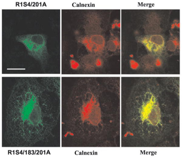
We carried out in vitro phosphorylation assays to determine whether ROMK1 is a substrate of protein kinase C (PKC) and used the two-electrode voltage clamp method to investigate the role of serine residues 4, 183, and 201, the three putative PKC phosphorylation sites, in the regulation of ROMK1 channel activity. Incubation of the purified His-tagged ROMK1 protein with PKC and radiolabeled ATP resulted in (32)P incorporation into ROMK1 detected by autoradiography. Moreover, the in vitro phosphorylation study of three synthesized peptides corresponding to amino acids 1-16, 174-189, and 196-211 of ROMK1 revealed that serine residues 4 and 201 of ROMK1 were the two main PKC phosphorylation sites. In contrast, (32)P incorporation of peptide 174-189 was absent. In vitro phosphorylation studies with ROMK1 mutants, R1S4/201A, R1S4/183A, and R1S183/201A, demonstrated that the phosphorylation levels of R1S4/201A were significantly lower than those of the other two mutants. Also, the Ba(2+)-sensitive K(+) current in oocytes injected with green fluorescent protein (GFP)-R1S4/201A was only 5% of that in oocytes injected with wild type GFP-ROMK1. In contrast, the K(+) current in oocytes injected with GFP-ROMK1 mutants containing either serine residue 4 or 201 was similar to those injected with wild type ROMK1. Confocal microscope imaging shows that the surface expression of the K(+) channels was significantly diminished in oocytes injected with R1S4/201A and completely absent in oocytes injected with R1S4/183/201A. Furthermore, the biotin labeling technique confirmed that the membrane fraction of ROMK channels was almost absent in HEK293 cells transfected with either R1S4/201A or R1S4/183/201A. However, when serine residues 4 and 201 were mutated to aspartate, the K(+) currents and the surface expression were completely restored. Finally, addition of calphostin C in the incubation medium significantly decreased the K(+) current in comparison with that under control conditions. Biotin labeling technique further indicated that inhibition of PKC decreases the surface ROMK1 expression in human embryonic kidney (HEK) cells transfected with ROMK1. We conclude that ROMK1 is a substrate of PKC and that serine residues 4 and 201 are the two main PKC phosphorylation sites that are essential for the expression of ROMK1 in the cell surface.


Figures

Fig. 1
Fig. 1
A, a Coomassie Blue staining shows that a 50-kDa protein was purified from E. coli transformed with ROMK1 mutants (R1S4A, R1S183A, R1S201A, R1S183/201A, R1S4/201A, and R1S4/183A). B, a Western blot analysis with ROMK antibody. IB, immunoblot.
Fig. 2
Fig. 2. An autoradiograph showing that radiolabeled ATP was incorporated into an 81- and a 50-kDa protein, respectively
Fig. 3
Fig. 3. Autoradiograph showing the PKC-induced phosphorylation of three synthesized peptides
A, an autoradiograph illustrating the results from an in vitro phosphorylation assay of three peptides corresponding to amino acid sequences of ROMK1, 196–211, 174–189, and 1–16, respectively. B, in vitro phosphorylation assay with peptides 1–16 and 1–16(S4A), which has the same sequence as that of 1–16 except that serine residue 4 was mutated to alanine. C, in vitro phosphorylation assay with peptides 196–211 and 196–211(S201A), which has the same sequence as that of peptide 196–211 except that serine residue 201 was mutated to alanine.
Fig. 4
Fig. 4. Autoradiograph demonstrating the PKC-mediated phosphorylation of ROMK1 R154/183A, R154/201A, and R15183/201A
In A, the top panel of the figure shows the result from an in vitro PKC phosphorylation of ROMK1 mutants, R1S4/183A, R1S4/201A and R1S183/201A. The bottom panel of the figure is a Coomassie Blue staining demonstrating the amount of proteins which were used to normalize data. The arrow indicates the position of ROMK1. In B, the top panel of the figure shows the result from an in vitro phosphorylation of ROMK1 and R1S183/201A. The bottom of the figure is a Coomassie Blue staining.
Fig. 5
Fig. 5. The Ba2+-sensitive K+ currents measured with two-electrode voltage clamp in oocytes injected with GFP-ROMK1 and its mutants, respectively
Each egg was injected with 25 ng of cRNA encoding ROMK1 or mutants.
Fig. 6
Fig. 6. Confocal microscope images demonstrating the surface expression of ROMK1 or mutants in oocytes injected with GFP-ROMK1 and ROMK1 mutants, respectively
The bar represents 100 μm.
Fig. 7
Fig. 7. Western blot showing membrane expression of ROMK1, R1S4/201A and R1S4/183/201A (A), and R1S4/201D (B)
The top panel demonstrates the biotin-labeled ROMK1 or mutants, and the bottom panel shows the total ROMK1 or mutant content in HEK293 cells. The ROMK1 position is indicated in the figure by a short bar. IB, immunoblot. IP, immunoprecipitation.
Fig. 8
Fig. 8. Confocal microscope images show the expression of ROMK1 and ROMK1 mutants in COS7 cells
Bar represents 25 μm.
Fig. 9
Fig. 9. Confocal microscope images demonstrate the location of R1S4/201A (green), R1S4/183/201A (green), and calnexin (red)
GFP-R1S4/201A and GFP-R1S4/183/201A were transiently transfected to COS7 cells. The cells were fixed with methanol 2 days after transfection, and the immunocytochemical study was carried out using antibodies of calnexin (1:100).
Fig. 10
Fig. 10. The effect of calphostin C (200 nm) on K+ current in oocytes injected with GFP-ROMK1 and GFP tagged R1S4/201D
After injection, oocytes were divided into control and experimental groups in which oocytes were incubated in a medium containing 200 nm calphostin C. K+ currents were measured 48–72 h after injection. The asterisk indicates that the data are significantly different from the control.
Fig. 11
Fig. 11. Western blot shows the effect of calphostin C on the biotin-labeled ROMK1 (A) and R1S4/201D (B)
HEK293 cells were transfected with GFP-ROMK1 or GFP-R1S4/201D in the presence or absence of calphostin C (200 nm). The surface K+ channels were labeled with biotin 48 h after transfection. The cell lysates were immunoprecipitated with GFP, and the biotin-labeled ROMK1/mutant was detected with avidin. The expression of ROMK1 or mutant in the cell membrane was normalized by comparison with its corresponding total ROMK channels. IB, immunoblot. IP, immunoprecipitation.

References

    1. Ho K, Nichols CG, Lederer WJ, Lytton J, Vassilev PM, Kanazirska MV, Hebert SC. Nature. 1993;362:31–38. - PubMed
    1. Wang WH, Hebert SC, Giebisch G. Annu Rev Physiol. 1997;59:413–436. - PubMed
    1. Xu JZ, Hall AE, Peterson LN, Bienkowski MJ, Eessalu TE, Hebert SC. Am J Physiol. 1997;273:F739–F748. - PubMed
    1. Mennitt PA, Wade JB, Ecelbarger CA, Palmer LG, Frindt G. J Am Soc Nephrol. 1997;8:1823–1830. - PubMed
    1. Giebisch G. Am J Physiol. 1998;274:F817–F833. - PubMed

Publication types

MeSH terms