. 2002 Sep;13(9):3148-61.
doi: 10.1091/mbc.01-11-0540.

The major sites of cellular phospholipid synthesis and molecular determinants of Fatty Acid and lipid head group specificity

Affiliations

The major sites of cellular phospholipid synthesis and molecular determinants of Fatty Acid and lipid head group specificity

Annette L Henneberry et al. Mol Biol Cell. 2002 Sep.

Abstract

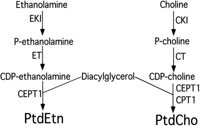
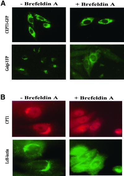
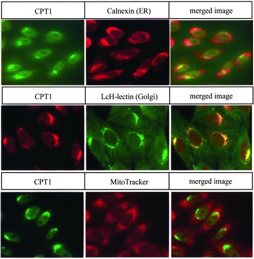
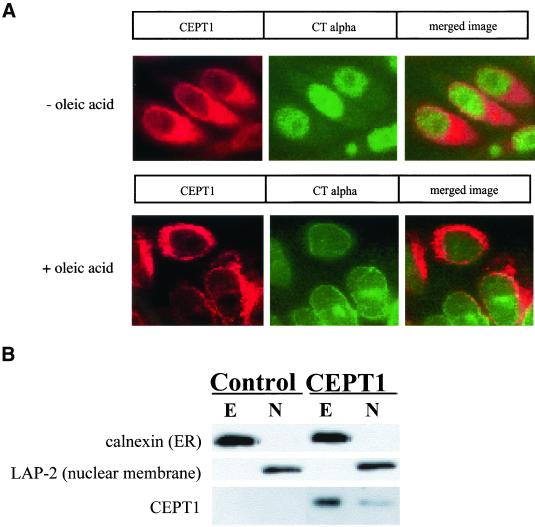
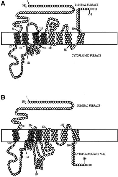
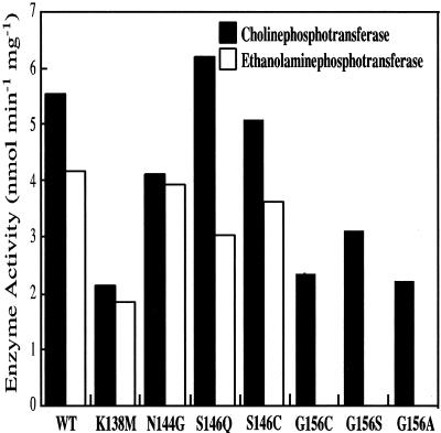
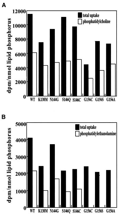
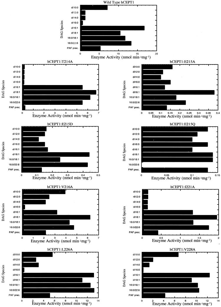
Phosphatidylcholine and phosphatidylethanolamine are the two main phospholipids in eukaryotic cells comprising ~50 and 25% of phospholipid mass, respectively. Phosphatidylcholine is synthesized almost exclusively through the CDP-choline pathway in essentially all mammalian cells. Phosphatidylethanolamine is synthesized through either the CDP-ethanolamine pathway or by the decarboxylation of phosphatidylserine, with the contribution of each pathway being cell type dependent. Two human genes, CEPT1 and CPT1, code for the total compliment of activities that directly synthesize phosphatidylcholine and phosphatidylethanolamine through the CDP-alcohol pathways. CEPT1 transfers a phosphobase from either CDP-choline or CDP-ethanolamine to diacylglycerol to synthesize both phosphatidylcholine and phosphatidylethanolamine, whereas CPT1 synthesizes phosphatidylcholine exclusively. We show through immunofluorescence that brefeldin A treatment relocalizes CPT1, but not CEPT1, implying CPT1 is found in the Golgi. A combination of coimmunofluorescence and subcellular fractionation experiments with various endoplasmic reticulum, Golgi, and nuclear markers confirmed that CPT1 was found in the Golgi and CEPT1 was found in both the endoplasmic reticulum and nuclear membranes. The rate-limiting step for phosphatidylcholine synthesis is catalyzed by the amphitropic CTP:phosphocholine cytidylyltransferase alpha, which is found in the nucleus in most cell types. CTP:phosphocholine cytidylyltransferase alpha is found immediately upstream cholinephosphotransferase, and it translocates from a soluble nuclear location to the nuclear membrane in response to activators of the CDP-choline pathway. Thus, substrate channeling of the CDP-choline produced by CTP:phosphocholine cytidylyltransferase alpha to nuclear located CEPT1 is the mechanism by which upregulation of the CDP-choline pathway increases de novo phosphatidylcholine biosynthesis. In addition, a series of CEPT1 site-directed mutants was generated that allowed for the assignment of specific amino acid residues as structural requirements that directly alter either phospholipid head group or fatty acyl composition. This pinpointed glycine 156 within the catalytic motif as being responsible for the dual CDP-alcohol specificity of CEPT1, whereas mutations within helix 214-228 allowed for the orientation of transmembrane helices surrounding the catalytic site to be definitively positioned.


Figures

Figure 1
Figure 1
Synthesis of PtdCho and PtdEtn by the CDP-alcohol pathways. The gene names of the enzymes that catalyze each step are indicated. EKI, ethanolamine kinase; ET CTP:phosphocholine cytidylyltransferase; CEPT1, choline/ethanolaminephosphotransferase; CKI, choline kinase; CT, CTP:phosphocholine cytidylyltransferase; CPT1, cholinephosphotransferase.
Figure 2
Figure 2
Effect of brefeldin A on CEPT1 and CPT1 subcellular localization. (A) CHO-K1 cells transiently transfected with T7-CEPT1-GFP or the first 81 amino of the Golgi resident β-1,4-galactosyltransferase fused to yellow fluorescent protein were treated with 2 μg/ml brefeldin A for 30 min. Live and fixed cells resulted in identical images. (B) CHO-K1 cells stably expressing T7-CPT1 were treated with brefeldin A and T7 monoclonal antibodies followed by Texas Red–conjugated secondary antibodies were used to determine the location of CPT1. The location of the Golgi was determined by the addition of FITC-coupled L. culinaris lectin.
Figure 3
Figure 3
Intracellular localization of CPT1. The intracellular location of CPT1 was determined in CHO-K1 cells stably expressing T7-CPT1 using T7 monoclonal primary antibodies. The location of the endoplasmic reticulum was assessed using primary antibodies to calnexin. Golgi location was determined by the addition of FITC-coupled L. culinaris lectin. MitoTracker dye was used to determine the location of the mitochondria. Secondary antibodies were coupled to either FITC or Texas Red. The yellow color in merged images indicates overlap between CPT1 and the organelle marker.
Figure 4
Figure 4
Intracellular localization of CEPT1. The intracellular location of CEPT1 was determined in CHO-K1 cells stably expressing T7-CEPT1 using T7 monoclonal primary antibodies. The location of the endoplasmic reticulum was assessed using primary antibodies to calnexin. Golgi location was determined by the addition of FITC coupled L. culinaris lectin. MitoTracker dye was used to determine the location of the mitochondria. Secondary antibodies were coupled to either FITC or Texas Red. The yellow color in merged images indicates overlap between CEPT1 and the organelle marker.
Figure 5
Figure 5
PtdCho synthesis pathway reconstitution at the nuclear membrane. (A) CHO-K1 stably expressing T7-CEPT1 were treated with oleic acid (500 μM in 0.5% bovine serum albumin) for 24 h to translocate CTα from its inactive soluble intranuclear location to its active nuclear membrane location before immunofluorescence. (B) Nuclei (N) were separated from extranuclear structures including the endoplasmic reticulum (E) by subcellular fractionation as described in MATERIALS AND METHODS, and the distribution of nuclear and endoplasmic reticulum markers are compared with CEPT1.
Figure 6
Figure 6
Predicted secondary structures for human CEPT1. Seven or eight membrane spans are strongly predicted with helix 181–199 positioned either in (A) or outside (B) of the membrane using the SMART or TmPred algorithms. The numbers denote amino acid residues with the black filled circles representing those mutated in the current study. Based on comparisons to studies on the yeast Cpt1p and Ept1p enzymes, the dark gray circles represent the maximum region required for diacylglycerol binding, and the light gray the maximum region required for CDP-alcohol binding. The conserved residues within the CDP-alcohol phosphotransferase motif, DG(x)2AR(x)8G(x)3D(x)3D, are also indicated.
Figure 7
Figure 7
CEPT1 CDP-alcohol phosphotransferase motif mutants. (A) The site-directed mutations made in CEPT1 are indicated. The numbers denote CEPT1 amino acid residues. (B) Western blot of wild-type and mutant versions of CEPT1.
Figure 8
Figure 8
Enzyme activity of CEPT1 CDP-alcohol phosphotransferase motif mutants. Enzyme activities were determined from microsomal membrane preparations of S. cerevisiae cells (cpt1:: LEU2 ept1) constitutively expressing CEPT1 or the indicated site-directed mutants. The ability to use either CDP-choline (black) or CDP-ethanolamine (white) as a substrate is indicated.
Figure 9
Figure 9
Metabolic reconstitution of PtdCho and PtdEtn synthesis by CEPT1 CDP-alcohol phosphotransferase mutants. Exponentially growing S. cerevisiae cells (cpt1:: LEU2 ept1) constitutively expressing CEPT1 or the indicated site-directed mutants were radiolabeled with (A) [14C]choline to label PtdCho or (B) [14C]ethanolamine to label PtdEtn, for 1 h. Radiolabel incorporated into each phospholipid was determined by scintillation counting.
Figure 10
Figure 10
CEPT1 diacylglycerol specificity mutants. (A) The site-directed mutations made in CEPT1 are indicated. The same region in human CPT1 is provided for comparison. The numbers denote CEPT1 amino acid residues. (B) Western blot of wild-type and mutant versions of CEPT1.
Figure 11
Figure 11
Enzyme activity of CEPT1 diacylglycerol specificity motif mutants. Enzyme activities were determined from microsomal membrane preparations of S. cerevisiae cells (cpt1:: LEU2 ept1) constitutively expressing CEPT1 or the indicated site-directed mutants.

References

    1. Ames BN, Dubin DT. The role of polyamines in the neutralization of bacteriophage deoxyribonucleic acid. J Biol Chem. 1960;235:769–775. - PubMed
    1. Attard GS, Templer RH, Smith WS, Hunt AN, Jackowski S. Modulation of CTP. phosphocholine cytidylyltransferase by membrane curvature elastic stress. Proc Natl Acad Sci USA. 2000;97:9032–9036. - PMC - PubMed
    1. Bae-Lee MS, Carman GM. Phosphatidylserine synthesis in Saccharomyces cerevisiae. Purification and characterization of membrane-associated phosphatidylserine synthase. J Biol Chem. 1984;259:10857–10862. - PubMed
    1. Bankaitis VA, Malehorn DE, Emr SD, Greene R. The Saccharomyces cerevisiae SEC14 gene encodes a cytosolic factor that is required for transport of secretory proteins from the yeast Golgi complex. J Cell Biol. 1989;108:1271–1281. - PMC - PubMed
    1. Bankaitis VA, Aitken JF, Cleves AE, Dowhan W. An essential role for a phospholipid transfer protein in yeast Golgi function. Nature. 1990;347:561–562. - PubMed

Publication types

MeSH terms

LinkOut - more resources