. 2002 Sep;13(9):3178-91.
doi: 10.1091/mbc.e02-05-0295.

Regulation of focal adhesion kinase by a novel protein inhibitor FIP200

Affiliations

Regulation of focal adhesion kinase by a novel protein inhibitor FIP200

Smita Abbi et al. Mol Biol Cell. 2002 Sep.

Abstract

Focal adhesion kinase (FAK) is a major mediator of integrin signaling pathways. The mechanisms of regulation of FAK activity and its associated cellular functions are not very well understood. Here, we present data suggesting that a novel protein FIP200 functions as an inhibitor for FAK. We show the association of endogenous FIP200 with FAK, which is decreased upon integrin-mediated cell adhesion concomitant with FAK activation. In vitro- and in vivo-binding studies indicate that FIP200 interacts with FAK through multiple domains directly. FIP200 bound to the kinase domain of FAK inhibited its kinase activity in vitro and its autophosphorylation in vivo. Overexpression of FIP200 or its segments inhibited cell spreading, cell migration, and cell cycle progression, which correlated with their inhibition of FAK activity in vivo. The inhibition of these cellular functions by FIP200 could be rescued by coexpression of FAK. Last, we show that disruption of the functional interaction between endogenous FIP200 with FAK leads to increased FAK phosphorylation and partial restoration of cell cycle progression in cells plated on poly-L-lysine, providing further support for FIP200 as a negative regulator of FAK. Together, these results identify FIP200 as a novel protein inhibitor for FAK.


Figures
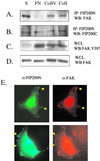
Figure 1
Figure 1
Association and localization of FIP200 and FAK. (A-D) Lysates were prepared from MDA-MB231 breast carcinoma cells that had been suspended, or replated on FN, type IV collagen, or type I collagen, as indicated. They were immunoprecipitated by anti-FIP200N and then analyzed by Western blotting with anti-FAK (A) or anti-FIP200C (B). The whole cell lysates (WCL) were also analyzed directly by Western blotting with anti-pFAKY397 (C) or anti-FAK (D). (E) NIH 3T3 cells (top panels) or ECV304 cells transfected with FIP200 (bottom panels) were processed for immunofluorescence as described in “Materials and Methods” using anti-FIP200N or anti-FAK, as indicated. Examples of colocalization of FIP200 and FAK in peripheral focal contacts are marked by arrows.
Figure 2
Figure 2
Analysis of FIP200 association with FAK. (A) A schematic of full-length FIP200 is shown on top. NT-FIP and CT-FIP segments, and MD-FIP of FIP200 are shown below. (B) 293T cells were transfected with an expression vector encoding HA-FAK and vectors encoding Flag-FIP200, its segments or empty vector control (V), as indicated. Lysates were immunoprecipitated with anti-Flag followed by Western blotting with anti-FAK to detect associated HA-FAK (top panel) or with anti-Flag to verify similar amounts of samples in the immunoprecipitates (bottom panel; Flag-FIP200 and segments are marked by arrowheads). (C) 293T cells were transfected with vectors encoding HA-FAK (WT) or its fragments (N-terminal, kinase domain and C-terminal) or with kinase domain of Pyk2, as indicated. Lysates from the transfected cells were then incubated with immobilized GST fusion proteins containing FIP200 segments or GST alone, as indicated. The bound proteins were resolved on SDS-PAGE and were analyzed by Western blotting with mAb 12CA5 (anti-HA). (D) Equal amounts of immobilized GST-fusion proteins containing FIP200 fragments, or GST alone, were incubated with 1 μg of recombinant FAK. The bound proteins were resolved on SDS-PAGE and were analyzed by Western blotting with anti-FAK (top panel). The membrane was also stained with Ponceau S stain to detect GST-fusion proteins (marked with arrowheads; bottom panel).
Figure 3
Figure 3
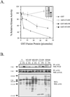
Inhibition of FAK activity by FIP200. (A) The kinase activity of FAK was assayed in the presence of various amounts of GST fusion proteins containing FIP200 segments, or GST alone, as indicated. Relative kinase activities were normalized to FAK activity in the absence of GST fusion protein. The mean ± SE of relative kinase activities from three independent experiments are shown. The inset shows Coomassie blue staining of a representation preparation of the fusion proteins (1, GST-NT-FIP; 2, GST-MD-FIP; and 3, GST-CT-FIP). (B) 293T cells were cotransfected with plasmid encoding HA-FAK and HA-FIP200, its segments, or vector control, as indicated. One day after transfection, cells were trypsinized and either kept in suspension (S) or replated on FN (10 μg/ml) for 30 min. They were then lysed and immunoprecipitated with anti-HA followed by Western blotting with PY20 to detect FAK phosphorylation (top panel) or anti-HA to verify similar FAK expression levels (middle panel). The corresponding whole cell lysates (WCL) were resolved on a SDS-PAGE gel and were western blotted with anti-HA to detect similar amounts of FIP200 and its fragments (marked by arrowheads, bottom panel).
Figure 4
Figure 4
Effects of FIP200 on FAK downstream signaling. 293 cells were cotransfected with plasmids encoding FAK, NT-FIP, or empty vector as controls, along with vectors encoding GFP-paxillin (A), Myc-p130cas (B), HA-Shc (C), or HA-Grb7 (D), as indicated. One day after transfection, cells were trypsinized and replated on FN (10 μg/ml) for 30 min. Whole cell lysates were then immunoprecipitated with anti-GFP, anti-Myc, or anti-HA to pull-down epitope-tagged paxillin, p130cas, Shc, and Grb7, respectively. The immune complexes were then analyzed by Western blotting with PY20 to detect their phosphorylation (top panels), or with antipaxillin, anti-Myc, or anti-HA to show their respective expression levels (bottom panels). The position of HA-Shc is indicated by arrows in C.
Figure 5
Figure 5
Inhibition of cell spreading by FIP200. (A) NIH3T3 cells were transfected with expression vectors encoding FIP200 or CT-FIP, as indicated. One day after transfection, cells were trypsinized and replated on FN (10 μg/ml) for 45 min. Cells were fixed and processed for immunostaining with anti-HA to detect the positively transfected cells (green) and with antivinculin to visualize all cells (red). (B-D) NIH3T3 cells were transfected with vectors encoding FIP200, its segments, or empty vector control, and plasmid encoding a HA-tagged FAK (in some experiments), along with a plasmid encoding β-Gal, as indicated. One day after transfection, cells were trypsinized and replated on FN (10 μg/ml) for 45 min. The cells were then fixed, and β-Gal assays were performed to identify the positively transfected cells. The mean ± SE of percentage of spread cells from three independent experiments are shown (B). Aliquots of whole cell lysates (WCL) were also analyzed directly by Western blotting using anti-Flag to detect FIP200 and its fragments (C) or anti-HA to detect FAK (D).
Figure 6
Figure 6
Inhibition of cell migration by FIP200. (A and B) NIH3T3 cells grown on FN (10 μg/ml) were transfected with FIP200, its segments, or empty vector control, with vectors encoding FAK or paxillin in some experiments, along with a plasmid encoding GFP in 7:1 ratio, as indicated. One day after transfection, the cell monolayer was wounded with a p10 tip, incubated at 37°C, and images were captured at 2-h intervals until 8 h. Images from representative experiments are shown in A. The rate of migration was measured by quantifying the total distance that the positively transfected cells (GFP+) moved from the edge of the wound toward the center of the wound in 8 h. (B) mean ± SE of the rate of migration from three independent experiments. *p = 0.48, 0,76, 0.32, and 0.89 for samples from left to right, in comparison with vector alone transfected cells. (C) Motility of cells on FN (5 μg/ml) were also assayed using OMAware based on time-lapse video microscopy as described in “Materials and Methods.” Representative field of cell tracks of control untransfected cells (a), cells transfected with expression vector encoding FIP200 (b), and cells transfected with both vectors encoding FIP200 and FAK (c) are shown. The arrows denote positively transfected cells. The untransfected cells in the same field serve as internal controls.
Figure 7
Figure 7
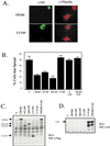
Regulation of cell cycle progression by FIP200. (A and B) NIH3T3 cells were transfected with expression vectors encoding HA-FIP200 or its segments, or an irrelevant control protein (C) or were mock transfected, as indicated. They were then analyzed for BrdU incorporation as described in “Materials and Methods.” (A) Representative fields for cells transfected with HA-FIP200 or the control. Immunostaining with anti-HA identifies positively transfected cells (green), and staining with anti-BrdU shows cells with new DNA synthesis (red). (B) The mean ± SE of three independent experiments of the percentage of BrdU+/positively transfected cells as determined by analyzing at least 80 positively transfected cells for each transfection in multiple fields. (C) NIH3T3 cells were cotransfected with an expression vector encoding FAK and that encoding HA-FIP200 or empty vector control (V), as indicated. A plasmid encoding β-Gal was also included. They were then analyzed for BrdU incorporation as described in “Materials and Methods.” The positively transfected cells were identified by immunostaining with anti-β-Gal. The percentage of BrdU+/β-Gal+ cells was determined by analyzing 40–50 β-Gal+ cells for each transfection in multiple fields. The percentage of BrdU-positive cells was normalized to the vector control of 100%. The results show mean ± SE of three independent experiments. *p = 0.18 and 0.59 are values for empty vector plus FAK transfection and HA-FIP200 plus FAK transfection, respectively, in comparison with value from empty vector alone transfection. Inset shows similar expression levels of FIP200 (α-HA blot) with or without cotransfection of FAK (KT3 blot).
Figure 8
Figure 8
Disruption of endogenous FIP200 interaction with FAK. (A and B) NIH3T3 cells were cotransfected with plasmid encoding HA-tagged KDKR or an irrelevant control protein (Grb7 SH2 domain, designated as C) and plasmids encoding HA-FAK or HA-Pyk2, as indicated. One day after transfection, cells were trypsinized and replated on PLL (0.1 mg/ml) or FN (10 μg/ml; A) or serum starved and treated with or without sorbitol (400 mM, 10 min; B), as indicated. Cell lysates were then immunoprecipitated with anti-HA or anti-Pyk2 and were western blotted with PY20 (top panel) or anti-HA (middle panel). Whole cell lysates (WCL) were also blotted with anti-HA (bottom panel) to show levels of transfected KDKR and control protein (C). (C) NIH3T3 cells were transfected with plasmid encoding HA-tagged KDKR or an irrelevant control protein, or mock transfected, as indicated. They were then analyzed for BrdU incorporation on both FN (10 μg/ml) and PLL (0.1 mg/ml) as described in “Materials and Methods.” The percentage of BrdU+/positively transfected cells was calculated and normalized to that of untransfected cells in each experiment. The mean ± SE are shown for data from three independent experiments.
Figure 9
Figure 9
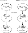
A working hypothesis of FIP200 interaction with FAK during cell adhesion.

References

    1. Angers-Loustau A, Cote JF, Charest A, Dowbenko D, Spencer S, Lasky LA, Tremblay ML. Protein tyrosine phosphatase-PEST regulates focal adhesion disassembly, migration, and cytokinesis in fibroblasts. J Cell Biol. 1999;144:1019–1031. - PMC - PubMed
    1. Arregui CO, Balsamo J, Lilien J. Impaired integrin-mediated adhesion and signaling in fibroblasts expressing a dominant-negative mutant PTP1B [published erratum appears in J. Cell Biol. 1998 143(6), 1761] J Cell Biol. 1998;143:861–873. - PMC - PubMed
    1. Avraham S, et al. Identification and characterization of a novel related adhesion focal tyrosine kinase (RAFTK) from megakaryocytes and brain. J Biol Chem. 1995;270:27742–27751. - PubMed
    1. Burridge K, Turner CE, Romer LH. Tyrosine phosphorylation of paxillin and pp125FAK accompanies cell adhesion to extracellular matrix: a role in cytoskeletal assembly. J Cell Biol. 1992;119:893–903. - PMC - PubMed
    1. Cary LA, Chang JF, Guan JL. Stimulation of cell migration by overexpression of focal adhesion kinase and its association with Src and Fyn. J Cell Sci. 1996;109:1787–1794. - PubMed

Publication types

MeSH terms