. 2002 Sep;13(9):3235-45.
doi: 10.1091/mbc.e02-02-0112.

Centrosomal proteins CG-NAP and kendrin provide microtubule nucleation sites by anchoring gamma-tubulin ring complex

Affiliations

Centrosomal proteins CG-NAP and kendrin provide microtubule nucleation sites by anchoring gamma-tubulin ring complex

Mikiko Takahashi et al. Mol Biol Cell. 2002 Sep.

Abstract

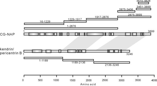
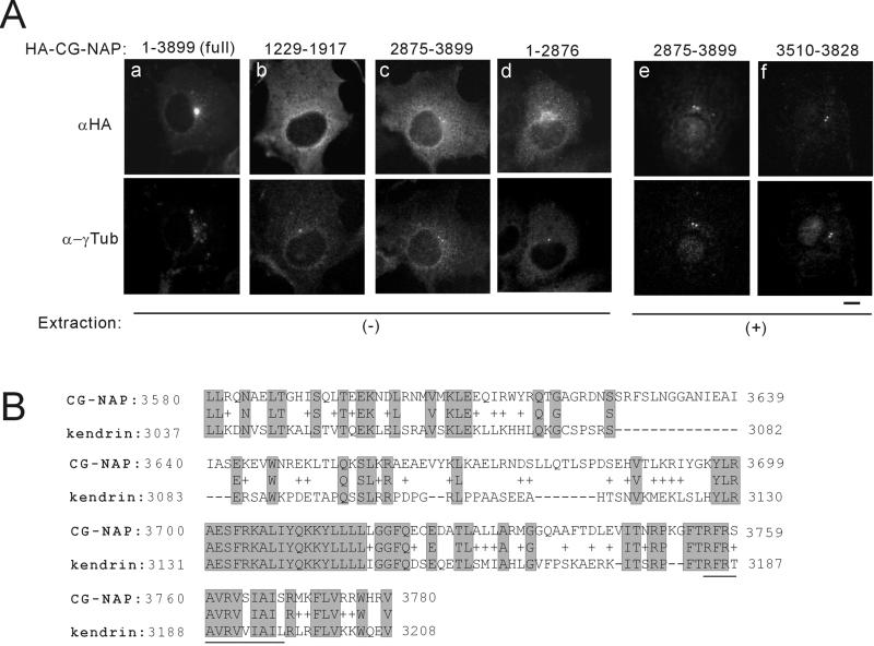
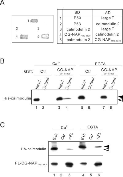
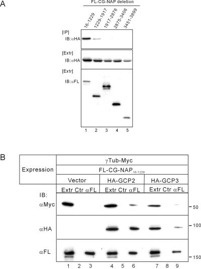
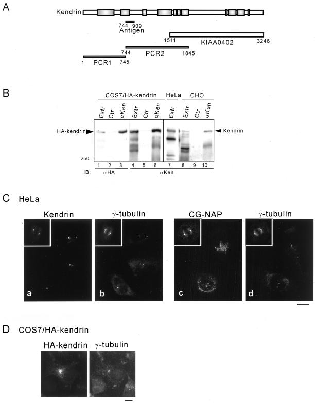
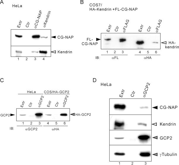
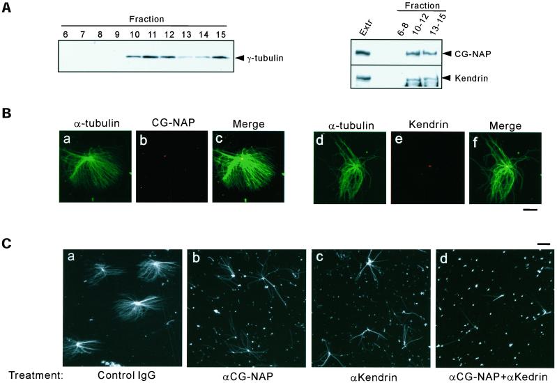
Microtubule assembly is initiated by the gamma-tubulin ring complex (gamma-TuRC). In yeast, the microtubule is nucleated from gamma-TuRC anchored to the amino-terminus of the spindle pole body component Spc110p, which interacts with calmodulin (Cmd1p) at the carboxy-terminus. However, mammalian protein that anchors gamma-TuRC remains to be elucidated. A giant coiled-coil protein, CG-NAP (centrosome and Golgi localized PKN-associated protein), was localized to the centrosome via the carboxyl-terminal region. This region was found to interact with calmodulin by yeast two-hybrid screening, and it shares high homology with the carboxyl-terminal region of another centrosomal coiled-coil protein, kendrin. The amino-terminal region of either CG-NAP or kendrin indirectly associated with gamma-tubulin through binding with gamma-tubulin complex protein 2 (GCP2) and/or GCP3. Furthermore, endogenous CG-NAP and kendrin were coimmunoprecipitated with each other and with endogenous GCP2 and gamma-tubulin, suggesting that CG-NAP and kendrin form complexes and interact with gamma-TuRC in vivo. These proteins were localized to the center of microtubule asters nucleated from isolated centrosomes. Pretreatment of the centrosomes by antibody to CG-NAP or kendrin moderately inhibited the microtubule nucleation; moreover, the combination of these antibodies resulted in stronger inhibition. These results imply that CG-NAP and kendrin provide sites for microtubule nucleation in the mammalian centrosome by anchoring gamma-TuRC.


Figures

Figure 1
Figure 1
Schematic representation of CG-NAP, kendrin, and their deletion mutants. Schematic structure of CG-NAP and kendrin are shown with predicted coiled-coil regions in shaded boxes. Positions of the deletion mutants of CG-NAP and kendrin are shown with amino acid residues on the upper and lower sides, respectively. Shaded areas between CG-NAP and kendrin represent the regions sharing homology found by BLAST search. Total amino acid residue of kendrin used in this study was 3246, as described in MATERIALS AND METHODS, which is shorter than that (3321) deposited to GenBank with accession number U52962.
Figure 2
Figure 2
Centrosomal localization of the carboxyl-terminal region of CG-NAP. (A) Subcellular localization of deletion mutants of CG-NAP. HA-tagged full-length and deletion mutants of CG-NAP were transiently expressed in COS7 cells, and then the cells were fixed with methanol directly (a–d) or after brief extraction with detergent to visualize proteins associated with intracellular structures (e, f). Then, the cells were double-stained with anti-HA and anti-γ-tubulin (α-γTub). Bar, 10 μm. (B) Sequence homology of the centrosomal-localization region of CG-NAP with kendrin. Aligned sequences are the result of BLAST search with CG-NAP3510–3828. The amino acid residues of kendrin shown are of U52962 and are different from those of our kendrin construct. Possible calmodulin binding sequence of kendrin homologous to that of yeast Spc110p (Flory et al., 2000) is underlined.
Figure 3
Figure 3
Association of the centrosomal-localization region CG-NAP3510–3828 with calmodulin. (A) Yeast two-hybrid analysis of interaction between CG-NAP3510–3828 and calmodulin 2. Interaction of the proteins fused to the Gal4 DNA binding domain (BD) and activation domain (AD) was assessed by growth and development of blue color of the transfected yeasts. Combinations of BD and AD constructs are shown on the right. p53 and SV40 large T antigen were used as controls. (B) Direct and Ca2+-dependent binding of CG-NAP3510–3828 with calmodulin. Bacterially expressed His6-tagged calmodulin 2 and GST-tagged CG-NAP3510–3828 were mixed and incubated in the presence of 2 mM CaCl2 or EGTA. After removal of aliquots (Input), glutathione-Sepharose beads were added to the mixture and incubated further, and then the proteins bound to the beads were collected (Output) and immunoblotted with anti-His. Black and white arrowheads indicate the positions of Ca2+-unbound and -bound forms of calmodulin, respectively. (C) Ca2+-independent coimmunoprecipitation of calmodulin with CG-NAP3510–3828. HA-tagged calmodulin 2 and FLAG-tagged CG-NAP3510–3828 were coexpressed in COS7 cells, and then cell extracts (Extr) were prepared in the presence of 2 mM CaCl2 or EGTA. The extracts were immunoprecipitated with anti-FLAG (αFL) or control (Ctr) mouse IgG followed by immunoblot with anti-HA (top) or anti-FLAG (bottom). Black and white arrowheads indicate the positions of calcium-unbound and -bound forms of calmodulin, respectively.
Figure 4
Figure 4
Association of CG-NAP16–1229 with γ-tubulin through binding with GCP2/GCP3. (A) Association of GCP2 with deletion mutants of CG-NAP. FLAG-tagged (FL) deletion mutants of CG-NAP were expressed in COS7 cells together with HA-tagged GCP2. Then the cell extracts were immunoprecipitated (IP) with anti-FLAG followed by immunoblot (IB) with anti-HA. (B) Indirect association of γ-tubulin (γTub) with CG-NAP16–1229 through binding with GCP2 or GCP3. COS7 cells were transfected with various combinations of expression plasmids as shown at the top. Then the cell extracts were immunoprecipitated with anti-FLAG or control mouse IgG followed by immunoblot with anti-Myc for γ-tubulin (top), anti-HA for GCP2 or GCP3 (middle), or anti-FLAG for CG-NAP16–1229 (bottom).
Figure 5
Figure 5
Centrosomal localization of endogenous and recombinant full-length kendrin. (A) Preparation of full-length cDNA of kendrin. Schematic representation of kendrin is shown with the fragments used to construct full-length cDNA as described in MATERIALS AND METHODS. Positions are shown with the corresponding amino acid residues. KIAA0402 was obtained from Kazusa DNA Research Institute. Position of the fragment to generate specific antibody is shown as Antigen. (B) Specificity of anti-kendrin (αKen) antibody. Cell extracts of COS7 cells expressing HA-tagged kendrin, HeLa, or CHO cells were immunoprecipitated with αKen followed by immunoblotting with anti-HA (lanes 1–3) or αKen (lanes 4–10). (C) Centrosomal localization of endogenous kendrin. HeLa cells were fixed with MeOH, and then double-stained with αKen and anti-γ-tubulin (a, b) or anti-CG-NAP (αEE) and anti-γ-tubulin (c, d). (D) Centrosomal localization of recombinant kendrin. COS7 cells expressing HA-tagged kendrin were fixed with MeOH, and then double-stained with anti-HA and anti-γ-tubulin. Bars, 10 μm.
Figure 6
Figure 6
Association of kendrin with γ-tubulin (γTub) through binding with GCP2. COS7 cells were transfected with various combinations of expression plasmids as shown at the top. Then the cell extracts (Extr) were immunoprecipitated with anti-FLAG (αFL) or control (Ctr) mouse IgG followed by immunoblot with anti-Myc for γ-tubulin (top), anti-HA for GCP2 or GCP3 (middle), or anti-FLAG for kendrin1–1189 (bottom).
Figure 7
Figure 7
Association of endogenous kendrin, CG-NAP, γ-tubulin, and GCP2. (A) Association of endogenous CG-NAP and kendrin. HeLa cell extracts (Extr) were immunoprecipitated with anti-CG-NAP (αEE+αBH) or anti-kendrin (αKen) or control (Ctr) rabbit IgG followed by immunoblot with anti-CG-NAP (αEE) (top) or αKen (bottom). (B) Association of recombinant kendrin with CG-NAP. 293T cells were cotransfected with FLAG-tagged (FL) CG-NAP and HA-tagged kendrin. Then the cell extracts were immunoprecipitated with anti-FLAG or control mouse IgG followed by immunoblot with anti-FLAG (lanes 1–3) or anti-HA (lanes 4–6). (C) Specificity of anti-GCP2 antibody. The extracts of COS7 cells transfected with HA-tagged GCP2 or HeLa cells were immunoprecipitated with αGCP2 or control rabbit IgG followed by immunoblot with αGCP2 (lanes 1–3) or anti-HA (lanes 4–6). (D) Association of GCP2 with CG-NAP and kendrin as well as with γ-tubulin. HeLa cell extracts were immunoprecipitated with αGCP2 or control rabbit IgG followed by immunoblot with anti-CG-NAP (αEE), αKen, αGCP2, or anti-γ-tubulin.
Figure 8
Figure 8
Inhibition of microtubule nucleation from isolated centrosomes by antibodies to CG-NAP and kendrin. (A) Presence of CG-NAP and kendrin in the fractions enriched with centrosomes. CHO cell lysates were fractionated by sucrose density gradient as described in MATERIALS AND METHODS. Then the fractions were examined for the presence of γ-tubulin (left), CG-NAP, and kendrin (right) by immunoblotting. Fractions 10–12 corresponding to the interphase between 40 and 60% sucrose were enriched with centrosomes. (B) Localization of CG-NAP and kendrin at the center of microtubule asters formed in vitro. Centrosome fractions were incubated with bovine tubulin in the presence of 1 mM GTP for 8 min at 37°C as described in MATERIALS AND METHODS. The resultant microtubule asters were fixed with glutaraldehyde and spun down on coverslips. Then the coverslips were processed for double-staining with anti-CG-NAP (αEE) and anti-α-tubulin (a–c), or αKen and anti-α-tubulin (d–f). Bar, 10 μm. (C) Inhibition of microtubule nucleation by pretreatment of the centrosomes with antibodies to CG-NAP and kendrin. Centrosome fractions were pretreated with antibodies to CG-NAP (αrXN) or kendrin (αKen) at a concentration of 1.2 mg/ml or with the combination of αrXN and αKen (1.2 mg/ml each), or control rabbit IgG (2.4 mg/ml) for 30 min on ice. Then microtubule nucleation was performed by incubation with bovine tubulin for 4 min. Microtubule asters were visualized by immunostaining with anti-α-tubulin. Bars, 10 μm. The results shown are representative of five different experiments.

Similar articles

Cited by

References

    1. Amano M, Mukai H, Ono Y, Chihara K, Matini T, Hamajima Y, Okawa K, Iwanatsu A, Kaibuchi K. Identification of aputative target at Rho as the serine-threonine kinase protein kinase N. Science. 1996;271:648–650. - PubMed
    1. Berchtold MW, Egli R, Rhyner JA, Hameister H, Strehler EE. Localization of the human bona fide calmodulin genes CALM1, CALM2, and CALM3 to chromosomes 14q24–q31, 2p21.1-p21.3, and 19q13.2-q13.3. Genomics. 1993;16:461–465. - PubMed
    1. Dictenberg JB, Zimmerman W, Sparks CA, Young A, Vidair C, Zheng Y, Carrington W, Fay FS, Doxsey SJ. Pericentrin and gamma-tubulin form a protein complex and are organized into a novel lattice at the centrosome. J Cell Biol. 1998;141:163–174. - PMC - PubMed
    1. Diviani D, Langeberg LK, Doxsey SJ, Scott JD. Pericentrin anchors protein kinase A at the centrosome through a newly identified RII-binding domain. Curr Biol. 2000;10:417–420. - PubMed
    1. Flory MR, Moser MJ, Monnat RJ, Jr, Davis TN. Identification of a human centrosomal calmodulin-binding protein that shares homology with pericentrin. Proc Natl Acad Sci USA. 2000;97:5919–5923. - PMC - PubMed

Publication types

MeSH terms