. 2002 Sep;13(9):3268-80.
doi: 10.1091/mbc.e02-03-0143.

A Rab8-specific GDP/GTP exchange factor is involved in actin remodeling and polarized membrane transport

Affiliations

A Rab8-specific GDP/GTP exchange factor is involved in actin remodeling and polarized membrane transport

Katarina Hattula et al. Mol Biol Cell. 2002 Sep.

Abstract

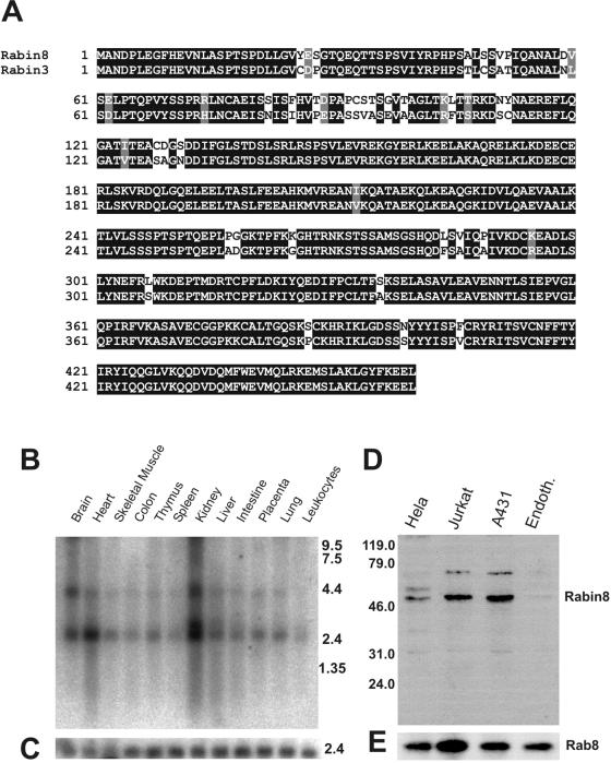
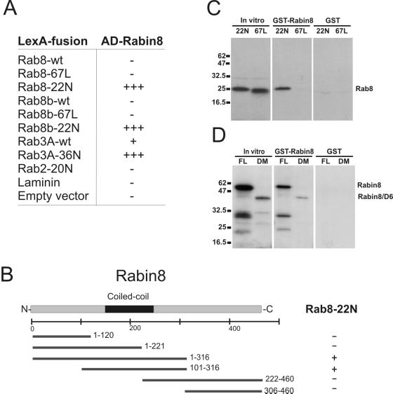
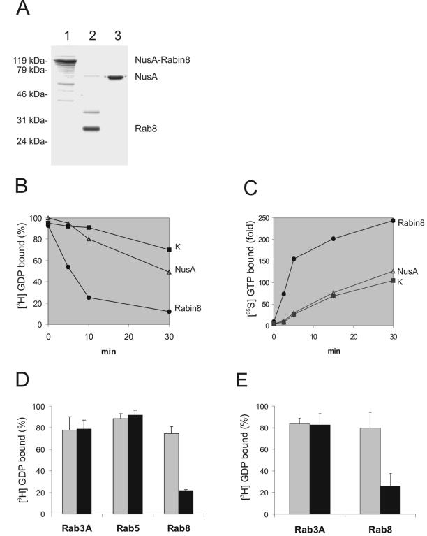
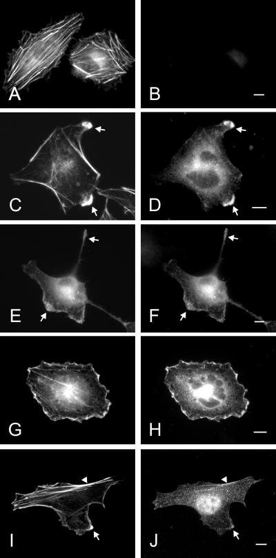
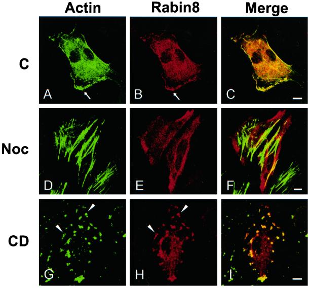
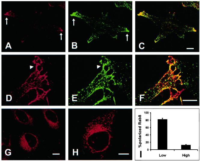
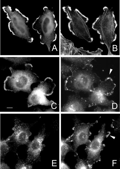
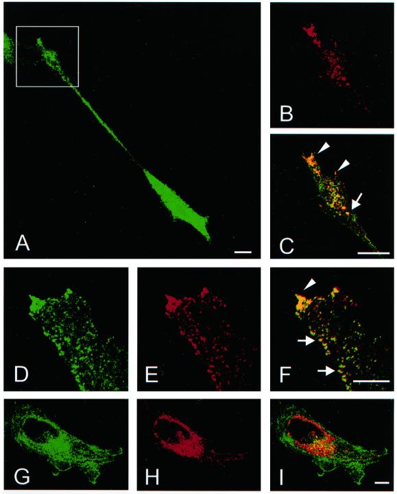
The mechanisms mediating polarized delivery of vesicles to cell surface domains are poorly understood in animal cells. We have previously shown that expression of Rab8 promotes the formation of new cell surface domains through reorganization of actin and microtubules. To unravel the function of Rab8, we used the yeast two-hybrid system to search for potential Rab8-specific activators. We identified a coil-coiled protein (Rabin8), homologous to the rat Rabin3 that stimulated nucleotide exchange on Rab8 but not on Rab3A and Rab5. Furthermore, we show that rat Rabin3 has exchange activity on Rab8 but not on Rab3A, supporting the view that rat Rabin3 is the rat equivalent of human Rabin8. Rabin8 localized to the cortical actin and expression of Rabin8 resulted in remodeling of actin and the formation of polarized cell surface domains. Activation of PKC by phorbol esters enhanced translocation of both Rabin8 and Rab8-specific vesicles to the outer edge of lamellipodial structures. Moreover, coexpression of Rabin8 with dominant negative Rab8 (T22N) redistributes Rabin8 from cortical actin to Rab8-specific vesicles and promotes their polarized transport to cell protrusions. The C-terminal region of Rabin8 plays an essential role in this transport. We propose that Rabin8 is a Rab8-specific activator that is connected to processes that mediate polarized membrane traffic to dynamic cell surface structures.


Figures

Figure 1
Figure 1
Amino acid sequence, tissue distribution, and endogenous Rabin8 protein in cell extracts. (A) Sequence alignment of human Rabin8 and rat Rabin3 obtained by ClustalW. Black boxes indicate identity and gray boxes indicate conservative amino acid substitutions (Boxshade software). (B) Northern blot analysis of Rabin8 RNA in different human tissues. (C) Actin RNA in corresponding tissues. (D) The presence of the endogenous Rabin8 protein in whole extracts of Hela, Jurkat, A431, and human endothelial cells was examined by Western blotting using affinity-purified anti-Rabin8. Molecular markers are indicated on the left. (E) Western blotting of same cell extracts detected by anti-Rab8.
Figure 2
Figure 2
Characterization of Rab8 binding to Rabin8. (A) Table showing interactions of Rabin8 in the two-hybrid system. The interactions were assayed by X-gal overlay and by the ability of the yeast to grow in the absence of leucine; −, no interaction; +, weak interaction; +++, strong interaction. (B) Schematic representation of Rabin8 deletions interacting with Rab8-T22N in yeast two-hybrid analysis. The black box indicates the coiled-coli region of Rabin8, and the numbers behind the bars indicate the amino acids encompassing the Rabin8 deletions. Positive interaction, +; negative interaction, −. (C) In vitro binding of Rab8 mutants to recombinant GST-Rabin8. Lanes beneath in vitro indicate input of translated Rab8-T22N and Rab8-Q67L, whereas lanes in the GST-Rabin8 column indicate in vitro bound Rab8-T22N and Rab8-Q67L to GST-Rabin8. The GST column shows the result obtained by GST alone. Molecular weight markers are indicated on left. (D) In vitro association of Rabin8 to GST-Rabin8. In vitro translated full-length (FL) Rabin8 or the Rabin8 1–316 deletion mutant (DM) seen as in put material in the in vitro column. The lanes beneath the GST-Rabin8 column indicate bound material of full-length (FL) and deleted Rabin8 (DM) to GST-Rabin. The column on right shows corresponding binding to the GST control beads. Positions of protein standards are given at the left of the gel.
Figure 3
Figure 3
Rabin8 and Rabin3 are nucleotide exchange factors for Rab8. (A) Recombinant NusA-Rabin8 (lane 1), Rab8 (lane 2), and NusA (lane 3) expressed and purified from E. coli lysates. Molecular markers seen at the left of the Coomassie brilliant blue–stained gel. (B) Rabin8 promotes dissociation of GDP from Rab8. Recombinant, wild-type Rab8 was loaded with [3H]GDP and then incubated alone (K), with NusA protein or with NusA-Rabin8. Aliquots of these three reactions were taken out at different time points and filtered on nitrocellulose filters. Note that Rab8 loaded [3H]GDP in the presence of NusA-Rabin8 lost 80% of its radioactivity within 10 min. (C) Rabin8 stimulates GTP association with Rab8. The [35S]GTP.γs binding to recombinant, wild-type Rab8 was measured after incubation at 37°C for indicated time periods with NusA-Rabin8, NusA, or Rab8 alone. The data are shown as the fold increase of [35S]GTP.γs binding compared with that obtained with nucleotide alone. (D) Rab3A, Rab5, and Rab8 were subjected to [3H]GDP release reactions in the presence (black bars) or absence (gray bars) of NusA-Rabin8. The percentage of [3H]GDP that remained bound to Rab3A, Rab5, and Rab8 after 20 min is presented. Values are means ± SEM from three independent experiments. (E) Rat Rabin3 is a Rab8-specific GEF. Purified NusA-Rabin3 was used to measure [3H]GDP release on Rab3A and Rab8. The percentage of [3H]GDP that remained bound to Rab3A and Rab8 after 20 min is presented. Values are means ± SEM from three independent experiments.
Figure 4
Figure 4
Overexpressed and endogenous Rabin8 in Hela cells. HeLa cells were transfected over night with empty vector (A and B) or with a construct containing a myc-tagged Rabin8 (C–H). Actin was localized by Alexa488-conjugated phalloidin (A, C, E, and G) and Rabin8 by anti-myc–specific mAb (B, D, F, and H). Protrusions, lamellae, and tails are indicated by arrows. Note that Rabin8-expressing cells contain fewer actin stress fibers and that the cells obtain ruffles, protrusions, and tails. Endogenous Rabin8 (J) and actin (I) were localized by affinity-purified anti-Rabin8 antibodies and Alexa488-conjugated phalloidin, respectively. Rabin8 staining along peripheral actin filaments are indicated by an arrowhead. Bars, 5 μm.
Figure 5
Figure 5
Rabin8 and the integrity of cytoskeletal elements. HeLa cells were transfected over night with a construct containing Rabin8. Then the cells were left untreated (A–C) or incubated next morning for 60 min with 1 μg/ml nocodazole (D–F) or for 20 min with 1 μM cytochalasin D (G–I). Analysis were done by confocal microscopy. Actin was detected by Alexa488-phalloidin (A, D, and G) and Rabin8 by an affinity-purified anti-Rabin8 antibody (B, E, and H). C, F, and I are corresponding merged pictures. Arrowheads indicate actin patches containing both actin and Rabin8 after cytochalasin D treatment. Note that there is no colocalization of actin with Rabin8 after nocodazole treatment. Bars, 5 μm.
Figure 6
Figure 6
Polarized distribution of endogenous Rab8 in HeLa cells. (A) In cells plated at low density, endogenous Rab8 is localized predominantly to the tip of protrusions (arrows), where Rab8 partially colocalize with the transferrin receptor (B and C). A strong localization of Rab8 to filopodia is often seen (D) where it colocalize with the transferrin receptor (E and F). In the cytoplasm the colocalization of Rab8 and the transferrin receptor is not often seen (F). In confluent monolayers Rab8 is localized to a perinuclear region (G). This perinuclear region is very similar to that seen for expressed Rab8–22N (H). (I) Polarized distribution of endogenous Rab8 was assessed quantitatively by comparing the staining of Rab8 in cells grown overnight at low density (low) or high density (high). All data is a mean ± SEM of three independent experiments (N = 50 cells). Bars, 5 μm.
Figure 7
Figure 7
Redistribution of Rab8 and Rabin8 after PKC activation. HeLa cells transiently transfected with Rabin8 were treated at 16 h with 100 ng/ml PMA for 30 min. Cells were fixed, permeabilized, and stained with alexa488-phalloidin to detect actin (B) and anti-Rabin8 (A). Similarly transfected cells were also used to detect both Rabin8 (C) and endogenous Rab8 (D) by anti-myc and anti-Rab8 antibodies. Untransfected cells treated with PMA for 30 min were used to compare tranferrin receptor (G) and endogenous Rab8 (H) distribution. Note that Rab8-specific vesicles obtain a peripheral distribution, whereas the transferrin receptor is localized more to vesicles in the cytoplasm. Arrowheads indicate Rab8-positive vesicles in the lamellipodia (D and F). Bar, 5 μm.
Figure 8
Figure 8
Rabin8 promotes polarized transport of Rab8-specific vesicles. HeLa cells were cotransfected with Rab8-T22N and myc-tagged Rabin8 cDNA and stained for Rab8 (A, D, and H) and myc (B, E, and G). Cotransfected cells exhibited protrusions (A) with growth cone like structures (square), with vesicles that contained both Rabin8 and Rab8-T22N (B and C). Numerous vesicular structures appeared in these cells (D, E, and F) that were seen to align along the cell periphery (arrows) and to accumulate in tips of protrusions (arrowhead). When cotransfection of Rab8–22N and a deletion mutant of Rabin8 (1–316aa) was done no polarization of Rab8-specific vesicles was seen (J, K, and I). In these cells Rab8–22N (H) remained in a perinuclear region and most of the Rabin8-deletion mutant in was found on the plasma membrane (G). However, there was partial colocalization of Rab8–22N and the Rabin8-deletion mutant in the perinuclear region (I). C, F, and I represent merge pictures. Bars, 5 μm.

References

    1. Armstrong J, Thompson N, Squire JH, Smith J, Hayes B, Solari R. Identification of a novel member of the Rab8 family from the rat basophilic leukemia cell line, RBL.2H3. J Cell Sci. 1996;109:1265–1274. - PubMed
    1. Beyaert R, Fiers W. Molecular mechanisms of tumor necrosis factor-induced cytotoxity: what we do understand and what we do not. FEBS Lett. 1994;340:9–16. - PubMed
    1. Bershadsky AD, Futerman AH. Disruption of the Golgi apparatus by brefeldin A blocks cell polarization and inhibits directed cell migration. Proc Natl Acad Sci USA. 1994;91:5686–5689. - PMC - PubMed
    1. Blangy A, Vignal E, Schmidt S, Debant A, Gauthier-Rouviere C, Fort P. TrioGEF1 controls Rac- and Cdc42-dependent cell structures through the direct activation of rhoG. J Cell Sci. 2000;113:729–739. - PubMed
    1. Bretscher MS. Getting membrane flow and the cytoskeleton to cooperate in moving cells. Cell. 1996;87:601–606. - PubMed

Publication types

MeSH terms

LinkOut - more resources